Amazing Facts About the Human Eye for Kids


In this Article
- What Is Human Eye?
- What Are the Different Layers of the Eye?
- What Are Parts of the Human Eye?
- How Does the Human Eye Work?
- Why Do We Have Two Eyes?
- Other Fun Facts and Information About Eyes for Children
View More
The human eye is an amazingly complex organ that uses different types of tissues to collect light and create vision. To understand how vision is produced, it is essential to understand how the lens in the eye changes shape to focus light onto the retina, where specialized cells sense the light. There are specialized structures that make up the eyeball that nourishes and maintains the different types of tissues, which contribute to the functioning of this organ. Children are often curious about how the eye works, and this article gives you a detailed yet simple explanation of how this organ works. Continue reading for important information and some great facts about your eyes.
What Is Human Eye?
The human eye is known as a sensory organ. It has evolved to collect light from our surroundings to produce vision. People have a pair of eyeballs which are spherical-shaped organs. They are located in the skull in bony sockets and face forward to create a 3-dimensional view of the world. The eyeballs contain a lens in the front that projects light onto the back of the eye called the retina. The retina has specialized cells that can detest brightness and color in the image formed by the lens. These cells transmit this information through the optic nerve to the brain. The brain deciphers the electrical impulses and interprets the image. The eyeball can be divided into three outer layers and the inner part, which is contained within the layers.
What Are the Different Layers of the Eye?
The eyeball is made up of 3 different layers:
1. Outer Coat
The outer coat comprises dense connective tissues called the sclera and cornea. These tissues are responsible for maintaining the shape of the eye and protecting it from damage.
2. Middle Coat
The middle layer comprises the choroid, ciliary body, and iris. Each has its function in contributing to the structure of the eyeball.
3. Inner Coat
The inner coat consists of the retina and its network of photoreceptors and nerve cells that transmit electrical impulses to the brain.
What Are Parts of the Human Eye?
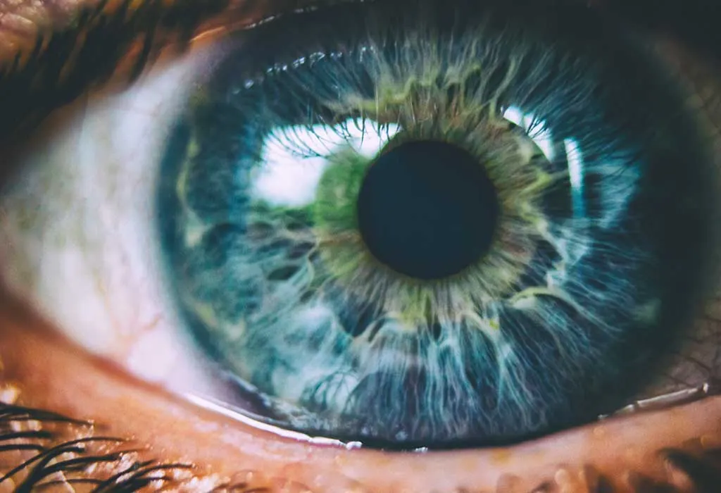
Anatomy of the Human Eye
When we look in the mirror, only a few parts of the eyes are visible. However, the eye is made up of different tissues and structures with specialized functions. Here are the important parts you need to know about:
1. The Sclera
The sclera is the distinctively visible white part of the eye. It is made out of opaque and fibrous tissue that acts as a protective layer for the eye. The sclera makes up most of the eyeball because it looks while throughout. It has a smooth and white appearance on the outside, while it is brown and grooved on the inside. The sclera and the dark iris on it enable us to identify a person’s direction of gaze.
2. Eyelids
The eyelids are foldable skin that acts as a protective cover for the eyes. Eyelids protect the eye from dust and debris, which can damage the delicate surface of the eyeball. The eyelids open and close when we blink our eyes or respond to a stimulus. They are also responsible for keeping the eye’s outer layer wet every time we blink or close our eyes.
3. Eyelashes
Eyelashes are the long hair that grows at the edge of the eyelids. Their main function is to catch dust and protect the eye from damage. The eyelashes allow us to see by squinting during high winds. It catches debris and allows us to see in conditions that would otherwise make us close our eyes.
4. Tear Glands
Each eye has a small tear gland, also called the lacrimal gland, at the corner of the eye. It secretes water, lacrimal gland fluid, proteins, and electrolytes. Each time we blink, the eyelids spread tears to moisten the eyes. Sometimes, excess tears flush out unwanted foreign particles such as dust, smoke, or other irritants.
5. Cornea
The cornea is the outermost layer in the front of the eye and borders the sclera. It is a transparent bulging surface that participates in vision by bending light.
6. Iris
The Iris is the dark and colored part of the eye in the middle of the white sclera that we all notice. It contains a sphincter muscle called the pupillary muscle that expands and contracts the iris to form a circular aperture called the pupil.
7. Pupil
The pupil is the eye’s part that appears as a black circle in the middle. It is a hole created by the Iris that can expand and contract to vary the eye’s aperture, thus controlling the amount of light entering the lens. When it is dark, the pupils expand to allow us to see more easily, and when it gets bright, it contracts to limit the light.
8. Lens
The lens is a transparent and flexible tissue located behind the iris. As the name suggests, it has the shape of a biconvex lens and can change its curvature to increase or decrease power to focus at different lengths. The lens is what bends the incoming light and forms the image on the retina.
9. Ciliary Body and Muscles
The ciliary body produces an aqueous humor fluid that fills up the anterior chamber in front of the lens. The ciliary muscles are attached to the lens and work to change the shape of the lens to focus at different distances.
10. Conjunctiva
The conjunctiva is a thin and clear membrane that covers the sclera and the inside of the eyelids. It protects the eye from microbes and keeps the eyeball lubricated by producing tears and mucus. While conjunctiva contributes to the formation of some tears, most of it is from the lacrimal glands.
11. Retina
The retina is a thin layer in the back that lines the inside of the eye. The focus light from the lens lands on the retina, where specialized cells detect light intensity and color and convert it into neural signals sent to the brain through the optic nerve. The specialized cells or photoreceptors are of two types:
- Rods: The rod cells enable us to see in low light conditions and are found in high concentration away from the retina’s center. Because of this, they also contribute to peripheral vision. The rod cells are also sensitive to movement, and each eye contains about 120 to 150 million rod cells.
- Cones: The cone cells are photoreceptors that enable color vision in daylight and bright light conditions. About 6-7 million cone cells are concentrated toward the retina’s center.
12. Optic Nerve
The optic nerve connects to the retina and transmits neural signals from the photoreceptors to the brain. It is also responsible for several neurological reflexes, such as the light and accommodation reflex. The optic nerve that connects to the retina is called a ‘blind spot’ as there are no photoreceptor cells in the region.
How Does the Human Eye Work?
Here is a step-by-step explanation of how vision is formed:
- Light rays from an object enter the eye through the cornea, slightly bent because of refraction. The cornea is the first step in the optical train that forms the image on the retina.
- The light from the cornea passes through the pupil. The Iris manages the amount of light that enters the eye by expanding or contracting the pupil.
- The light then passes through the double convex eye lens to form an image on the retina. The lens and the cornea together form a sharp image on the retina in people with normal vision.
- The image formed on the retina is sensed by the photoreceptors and turned into electrical signals that travel to the brain via the optic nerve.
- The brain then turns the electrical signals into imagery that we perceive.
Why Do We Have Two Eyes?
We have binocular vision in the two sets of eyes, which is essential for depth perception. Since our eyes are set apart, they can view the same object from different angles and form a three-dimensional image in the brain. This ‘convergence’ in the brain helps us perceive distances, sizes, and depth of things away from us.
Other Fun Facts and Information About Eyes for Children
Here are some eye fun facts that show how amazing this structure is:
- Your eye can distinguish about 10 million different colors.
- The brain is the only organ more complex than the eye.
- When it comes to weird facts about eyes, a mention should be given to heterochromia. It is a condition in which the irises of both eyes have different colors. It can be caused by an injury or at birth.
- People who have farsightedness have shorter eyeballs. People who have near-sightedness have longer eyeballs than average.
- Brown is the most common eye color in the world.
- It is not possible to sneeze with your eyes open!
The human eye is the most complex organ other than the brain. When you look at some of the cool facts about eyes, it becomes evident that this is an amazing organ essential for the human body.
Also Read:
Plants Facts for Children
Science Facts for Children
Facts about Funny Bone for Kids
Was This Article Helpful?
Parenting is a huge responsibility, for you as a caregiver, but also for us as a parenting content platform. We understand that and take our responsibility of creating credible content seriously. FirstCry Parenting articles are written and published only after extensive research using factually sound references to deliver quality content that is accurate, validated by experts, and completely reliable. To understand how we go about creating content that is credible, read our editorial policy here.
- Author
Related Articles
-

Essay on Onam for Students & Children in English
-

15 Must-Know Pollution Facts for Kids
-

Speech on Father's Day in English for Children & Students
-

Teaching Size and Shapes to Preschoolers
-

These 7 Tips Will Help Your Child Survive Maths - Whether They Love it or Hate it!
-

10 Tips on Tutoring Your Children Yourself at Home

.svg)


")


