

















.svg)
.svg)
.png)


Pregnancy Week By Week
Suggested articles for you
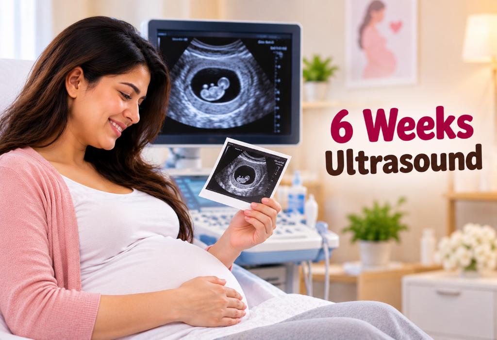
6 Weeks Ultrasound: Procedure, Abnormalities and more
Key Takeaways
A 6-week ultrasound can help confirm the pregnancy’s location, estimate gestational age, and assess early development.
It may be possible to see the gestational sac, yolk sac, embryo, and cardiac activity, depending on pregnancy dating.
Not seeing an embryo or heartbeat at 6 weeks does not always indicate a problem; a repeat scan may be recommended.
A six weeks ultrasound can help confirm the location and early development of a pregnancy, although what is visible at this stage can vary. The World Health Organization (WHO) recommends that pregnant women have at least one ultrasound before 24 weeks to help estimate gestational age and improve the detection of fetal anomalies and multiple pregnancies (1).
At six weeks, it is natural to wonder what the scan should show and whether an unclear finding means something is wrong. This article explains the procedure, what doctors look for, possible abnormalities, and when a repeat scan may be advised.
Why Is a 6-Week Pregnancy Ultrasound Scan Required?
There are a few reasons why an ultrasound scan is required during the 6th week of pregnancy (2):
To rule out ectopic pregnancy and confirm that the pregnancy sac is located inside the uterus.
To check if the heart of the foetus is beating.
To calculate the number of weeks of pregnancy or the gestation period that have passed.
To detect any bleeding or spotting in the internal organs.
To check the number of babies in the womb.
To confirm if there are complications in the
To check the mother’s pelvic organs.
Preparations Required for a Six-Week Pregnancy Scan
An ultrasound for pregnancy at 6 weeks generally does not require any special preparation. However, a few simple steps beforehand can help ensure clearer images:
Your technician may ask you to drink plenty of water beforehand to keep your bladder full.
Drinking around four to five glasses of water about an hour before the scan is generally advised.
Try not to urinate before the scan unless your healthcare provider advises otherwise.
How Long Does a Six-Week Ultrasound Take?
The scanning process does not take a lot of time. The entire process is usually over within a few minutes; it will seldom take more than half an hour.
How Is the Scan Performed?
As mentioned earlier, there are two ways to carry out an ultrasound scan. One is the transabdominal scan, and the other is the transvaginal scan. For both the scans, the expecting mother is made to lie on her back on a bed. During a transabdominal scan, her tummy is lubricated using a gel, and a scanning probe is pressed on top of her belly in a firm manner (3). During a transvaginal scan, a thinner scanning probe is inserted in the vagina. In both cases, the images are studied on a computer screen (4).
Usually, the quality of the images obtained during a transabdominal scan helps the technician determine whether you need a transvaginal ultrasound scan. That’s because transvaginal ultrasound scans provide more clear images (5). The probes used for both ultrasound scans emit ultrasonic sound waves that help capture an image of the baby inside the womb. These images are then evaluated by the doctor for assessing your pregnancy.
What Is Assessed Through an Ultrasound Scan?
Many mothers wonder what exactly to expect in an ultrasound scan done in the 6th week. Well, several aspects of the pregnancy and the baby in the womb can be obtained using the scanning procedure. Here are some of the developments or what you can see in ultrasound at 6 weeks (6):
The location of the pregnancy– whether it is ectopic or within the uterus – is obtained.
You can get an idea of whether you will be able to carry the pregnancy to full term.
You can also obtain the foetal pole of the baby, which is the shape of the embryo as it develops gradually in the uterus. The head area and the leg buds are usually visible and can be differentiated by the technician.
The chorion and yolk sacs, both of which are absolutely essential for the baby to grow healthily, can be clearly seen in an ultrasound scan. The sacs provide the nutrition required by the embryo until the placenta has been established.
The heartbeat of the baby is another aspect commonly detected, although it grows stronger around the seven-week mark.
How Does a Baby Look Like at 6 Weeks of Pregnancy?
At six weeks of gestation, the embryo becomes visible as a featureless oval structure next to a yolk ac, which initially measures 1-2 mm in length. The foetus at this stage of pregnancy is similar to the size of a single sweet pea, about 3/4 inch in length (7). The little blip or baby’s back is curved along with a large head.
Is It Possible To See Twins in the 6-Week Ultrasound?
Yes, it is possible to see twins at 6 weeks ultrasound. The ultrasound can reveal the separate yolk sacs and their heartbeat. The twin ultrasound at 6 weeks will show two dark spots as babies in the uterus.
Does a 6-Week Ultrasound Scan Fail to Reveal the Embryo?
In some cases, the 6 weeks scan may not reveal the embryo or any of its vital signs. The lack of a heartbeat does not mean anything significant, as later scans are sure to detect the heartbeat of the foetus. You might be off about the timing of the pregnancy, thinking that it has gone on for longer than it actually has. This usually means that the signs of the embryo come along later than you thought they would.
Pictures of 6-Week Ultrasound
At a six-week ultrasound, the foetus will be the size of a sweet pea. It is quite normal for only the yolk sac to be visible during this early phase. There is no need to be alarmed if you see only a small, seemingly empty, dark circle on the ultrasound.
Since it’s still quite early in the pregnancy journey, doctors often assess the diameter of the gestational sac as a measurement. Typically, around the 6th week of pregnancy, the gestational sac’s diameter is approximately 10 millimetres.
6-Week Ultrasound and No Heartbeat – Is There a Chance of Miscarriage?
Although a 6-week ultrasound 3D scan can give the doctor an idea about the chances of a miscarriage, the fact that a heartbeat was not detected is not a sign of an impending tragedy. The heartbeat might simply be too faint, and it can be detected after a week or two when it has grown sufficiently. If you experience heavy bleeding or period cramps, then checking for a miscarriage at 6 weeks pregnant ultrasound can be beneficial.
What If Any Other Abnormalities Are Found on the Scan?
The first pregnancy scan done in the 6th week of pregnancy gives the gynaecologist a fair evaluation of how the pregnancy is progressing and an idea of whether there are growth abnormalities in the foetus. If there are likely any abnormalities, the doctor will surely help you tackle the problem and ensure the best for your baby with the help of a 6-week foetus ultrasound.
FAQs
1. Is a transvaginal ultrasound needed at 6 weeks?
A transvaginal ultrasound may be recommended because it can provide clearer images of an early pregnancy than an abdominal scan. It involves gently inserting a covered ultrasound probe into the vagina and is generally considered safe during early pregnancy.
2. Can a 6-week ultrasound detect an ectopic pregnancy?
An early ultrasound can help determine whether the pregnancy is developing inside the uterus. If a pregnancy is not seen within the uterus or there are concerning findings, the doctor may investigate the possibility of an ectopic pregnancy using ultrasound and blood tests.
3. Can the baby’s gender be determined at 6 weeks?
No. A 6-week ultrasound is far too early to reliably determine the baby’s sex. The scan primarily focuses on confirming and assessing early pregnancy development.
4. Can a 6-week ultrasound show the baby’s movements?
The embryo is extremely small at 6 weeks, so noticeable movements are generally not expected. The main focus of the scan is on early structures and, when visible, cardiac activity.
An ultrasound scan at 6 weeks is a must for any pregnant woman, not just to evaluate the health of the child but that of the mother too. It also helps decrease anxiety and helps take necessary and prompt actions in case of abnormalities.
Previous Week: 5th Weeks Pregnancy Ultrasound
Next Week: 7th Weeks Pregnancy Ultrasound Scan
Read more




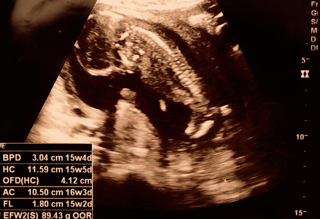
15 Week Ultrasound – What to Expect?
Key Takeaways
A 15-week ultrasound can check your baby’s growth, movements, heartbeat, and developing anatomy.
Most scans at this stage are performed through the abdomen and are generally quick and non-invasive.
Your baby is around 10 cm long and weighs about 70 g at 15 weeks, although measurements can vary.
A 15-week scan can show movements and developing features, but it cannot detect every possible abnormality.
A 15th week of pregnancy ultrasound can give you a closer look at your baby’s growth, movements, heartbeat, and developing anatomy. At this stage, the scan may also help your healthcare provider assess gestational age and check the baby’s overall development. The World Health Organization (WHO) recommends at least one ultrasound before 24 weeks of pregnancy (1).
As your baby continues to grow and develop, you may be curious about what you’ll actually see during the scan and what the images can tell you. This article explains how a 15-week ultrasound is performed, what you may see during the scan, how your baby is developing, and what questions you may want to ask your doctor.
Why You Need 15 Week Ultrasound?
There are many reasons why a mother needs to know about the growth of her baby during the second trimester. Here is why you need a 15 week fetus ultrasound (2) (3).
Doctors do this scan to see if the baby is growing appropriately.
You can see how much the baby has grown by this time.
You might get to see a few expressions on the baby’s face. Nothing makes a mother happier than seeing her child smile (or frown) for the first time.
How to Prepare for the Scan?
Preparing for 15 week sonogram does not include anything considerably different, compared to the earlier tests. Like in all ultrasound scans, you will be encouraged to drink a lot of water beforehand. This ensures that you have a full bladder, which will help the technician to get clear images of the child. So you can start preparing by drinking at least a litre (or more) of water 1-2 hours before the process is to begin so that you have the urge to urinate at the time of the scan. Also, you should not urinate again until after the 15 week scan is complete.
15-Week Ultrasound – How Long It Takes to Complete the Scan?
The scanning process is usually done within ten to fifteen minutes, as all ultrasound scans do not take much time. If there are any technical malfunctions, the process may take longer. However, in most cases, the test gets done within half an hour or so.
15-Week Ultrasound – How It Is Performed?
Usually, the sonogram at 15 weeks pregnant do not include any invasive procedures. You will be made to lie on your back, with your belly showing. The technician will apply jelly all over the stomach, for reducing friction while moving the probe. The probe is then pressed on top of your belly and moved around to get real-time images on a computer screen. The technician takes snapshots when he/she gets some images (4). The entire process does not last long unless the images are not clear enough. In such cases, a plastic probe emitting ultrasound waves will be inserted into your vagina, so that the technician can get clear images of the child (5). In all cases, the test does not take more than half an hour to complete.
15-Week Ultrasound – What Can Be Seen?
The images of a baby at 15 weeks ultrasound scan can tell you and the doctor a lot about how the baby has been growing. You will be able to watch your child make a lot of funny expressions during the scan – the baby will grimace, frown, smile, and even squint. This is just the child trying out his new, developing muscles. You might even be able to see the child swallowing or sucking his thumb (6).
Arms and legs could be moving, too. The images will show the skin of the child, which will have a paper-like consistency. The blood vessels running underneath will be visible. The scalp of the child, along with the eyebrows is also visible at this stage. Your child may already have hair on his head. This temporary, fine hair is called the lanugo (7).
The skeletal system of the child is still developing, as the cartilage in place slowly transforms into the harder bone (8). However, worry not; the bones still stay flexible enough so that the child can pass through the birth canal without difficulty. They do not harden enough for the child to stand until he is a toddler. He might also be able to turn his head and make fists at this time. The development of external genitalia will also be present, so the doctor will be able to guess the gender of the baby with almost 80% accuracy.
How Big Is Your Baby at 15 Weeks?
At 15 weeks pregnant, your baby is about 10 cm (4 inches) long from head to bottom and weighs around 70 grams, roughly the size of an orange (9). Your baby is also becoming more active, although you may not feel those movements yet.
15-Week Ultrasound – Identifying Abnormalities
The 15-week foetus ultrasound scan definitely gives the doctor a clear idea about how the baby has been growing, so he might advise you to have further tests if he detects anything out of place. Although the 3D ultrasound at 15 weeks is quite comprehensive, it is still not enough to be sure.
There are no routine tests scheduled for mothers at this time of pregnancy, but women above the age of 35, who are pregnant may benefit from doing an amniocentesis and multiple marker screening tests (10). The former uses a sample of amniotic fluid from your womb, in order to check for genetic abnormalities in the child.
FAQs
1. Can I hear the heartbeat of my baby at the 15 week ultrasound?
Yes. On average, you should be able to hear the heartbeat of your baby between 12 -14 weeks. In some cases it might take longer, but your doctor will check and advise.
2. What is the 15 week ultrasound called?
The medical term for a fifteen week ultrasound is a mid-pregnancy transabdominal ultrasound, as this is the approximate time that the second trimester begins.
3. Can you see the baby’s gender at 15 weeks?
Sometimes, the baby’s sex may be visible at 15 weeks if the position is suitable and the ultrasound equipment provides a clear view. However, it may not always be possible to determine it accurately at this stage.
4. Can you have a 4D ultrasound at 15 weeks?
A 4D ultrasound can sometimes be performed at 15 weeks, although the images may not be as detailed as those obtained later in pregnancy. Your baby’s position and the amount of amniotic fluid can also affect how clearly the face and body are seen.
An ultrasound scan at the 15th week of pregnancy can really benefit both the mother and the child. The doctor can see how well the child has been growing, while the mother gets to see the child make expressions for the first time. It can also help mothers detect any sort of abnormality in the development of the child at an early stage, so the scan at this time is a highly recommended one.
Previous Week: 14 Weeks Pregnant Ultrasound
Next Week: 16 Weeks Pregnant Ultrasound
Read more

2 Weeks Pregnant – Symptoms, Baby Size & Care Tips
If you’re 2 weeks pregnant, you may be surprised to learn that you’re probably not pregnant yet. Instead, your body is preparing for ovulation, with conception typically occurring around the end of this week. This means you may not have noticeable pregnancy symptoms or a measurable baby size just yet. Still, it’s an important time to start pregnancy-friendly habits and give your body the nutrients and care it needs.
In this article, we’ll explain what happens at two weeks pregnant, when conception occurs, what “baby size” means at this stage, possible sign and symptoms of two weeks pregnancy, and practical care tips, including folic acid and healthy lifestyle choices.
Your Baby During Week 2 of Pregnancy
Welcome to the beginning. That’s right, you’re about to the start the most exciting journey any human has ever had, and it’s going to be filled with thrills, chills, tears and laughter. Every moment is going to soon become the most memorable, and yet the next one will surprise you by surpassing the last.
The second week of pregnancy is when you may be about to release an egg that could eventually be fertilised by a sperm. This means you are not pregnant yet. Confused? Let’s understand this more clearly.
Your due date is calculated from the first day of your last menstrual period (1). So, in the second week, you are in the phase where you are most likely to get pregnant if you have been trying. At the 2 week mark, your egg has been released from the ovary and is ready to be fertilised. Your uterus has thickened to accept the fertilised egg (2).
The exact day of ovulation is hard to predict and may be anytime from the 9th day to the 21st day (3). The egg is fertilised 24 hours after the egg is released by one of the sperms that manage to swim up the fallopian tube. Only about a few hundred sperms manage to reach the fallopian tube from the millions released during ejaculation. About 10-30 hours after fertilisation, the nucleus of the sperm and the egg from the zygote fuse, deciding if you are going to have a boy or a girl (if the sperm has an x chromosome, your baby is a girl, and it has a y chromosome, your baby is a boy).
In the next 3 to 4 days, the zygote divides into 16 cells, and when it reaches the uterus, it is called the morula. The morula, a little ball of cells, burrows into the uterine lining and begins the process of division into the 2 week embryo and the placenta (4).
It has been 2 weeks since your menstrual period ended, and for some, the excitement is about to start. If you have not been trying to get pregnant, you may even miss out on recognising the early pregnancy symptoms, and brush them off as PMS or a late period.
What Is the Size of the Baby at Week 2 of Pregnancy?
Many women do not realise that at 2 weeks of pregnancy, the egg is not yet fertilised. At best, you are ovulating. We say, at best, because on day 15 of your menstrual period, you should be ovulating but only if you are one of the lucky few who has a 28-day menstrual cycle. If not, then you probably are a few days behind on ovulation. Many women are misinformed about what it means to be 2 weeks pregnant. The 2 week fetus size, health and development are still a good 2 to 3 weeks away.
As the pregnancy progresses, your body will go through some changes too. Let’s learn what those changes will be.
Common Body Changes at Week 2 of Pregnancy
There are a host of different ways your body will change during pregnancy. Some changes like larger breasts are universal, while other body changes during pregnancy like a change in hair texture are different for different women.
The beginning of the second week is when your body produces the Follicle Simulating Hormone, a hormone which is responsible for helping the dominant follicle mature and rupture. A ruptured follicle is called the corpus luteum, and it produces progesterone and oestrogen. These are also called pregnancy hormones are not only responsible for sustaining the pregnancy, but are also the cause of the symptoms you may already have started experiencing (5).
At the two-week pregnancy mark, since there is no actual fertilisation, you will experience symptoms exactly similar to those you would have during your menstrual period. This will include some breast soreness, light pelvic ache and even an increased sex drive.
2 Weeks Pregnant Symptoms
At the 2-week mark of your menstrual period, you should be ideally ovulating. If you are looking to get pregnant and would like to know the signs of when you could conceive, then you have come to the right place. Take a look at these symptoms in week 2 of pregnancy (6):
White cervical mucus: The mucus lining of your cervix changes to allow for the sperm to travel better. This is the reason for the whitish discharge from your vagina at this time.
Increased sense of smell: Like most other pregnancy symptoms, this one can be blamed in oestrogen too, and is usually what triggers morning sickness.
Light spotting: When the follicle around the egg ruptures, you might see some light spotting. However, if the bleeding is heavier, it could be indicative of a more serious condition. Consult with a doctor if you are worried about any of these symptoms.
BBT: The first two weeks of pregnancy are also called the follicular phase. If you have been tracking your basal body temperature (BBT) , you will notice that it is low at this stage, and starts to rise as you enter the luteal phase.
These symptoms should give you a good indication of when you are ovulating. You can also use a journal to track your period if it isn’t a 28-day cycle. There are also ovulation kits and fertility monitors available in the market for you to zero in on an ideal time to conceive. Once you conceive, you should keep a lookout for signs of pregnancy and immediately talk to your doctor.
Belly at Week 2 of Pregnancy
The fertilised egg has not yet attached itself to your uterine wall. In this condition, you will not see any changes in your 2 week pregnancy belly. Some women do complain of pelvic pain during ovulation, and that is probably the most you will feel during this time.
Ultrasound at Week 2 of Pregnancy
Pregnancy tests can be reliable only at the 4-week mark. This is the reason ultrasound is not done during the two-week mark. If you were to see your reproductive organs during this time, you would see a tiny egg travelling down the fallopian tube to be fertilised by sperm. However, the egg is smaller than a grain of salt, and you will probably not see anything. At best, the technician will be able to tell if there is a thickening of the uterine wall.
Diet at Week 2 of Pregnancy
Eating a healthy diet always goes a long way in making sure that your body is healthy enough for optimum body functioning, including getting pregnant. However, apart from the regular balanced diet, you do not have any special dietary needs as far as the 2nd week of pregnancy food is concerned.
Making healthy meal choices in early pregnancy goes a long way in ensuring that your baby develops well, and your body is able to meet the demands childbirth puts on it. If you are eager to conceive, try to include fertility-boosting foods such as leafy greens, figs, salmon, yams, and different kinds of berries. Try and reduce the intake of caffeine as it is believed to lower the chances of conception. It is a good idea to pick foods high in folic acid such as spinach and speak to your doctor about a folic acid supplement, too. This will ensure that the chances of birth defects, including neural tube defects (birth defects of the spinal cord), are reduced (7).
Week 2 of Pregnancy – Tips & Care
Taking care of yourself during this fun-filled, exciting period can be hard and confusing because of the immense number of research materials out on the internet. Remember to talk to your OB/GYN before implementing any care. Here are a few things you should keep in mind during the 2nd week of your pregnancy.
Dos
Keep track of your menstrual cycle.
Use an ovulation kit.
Have sex every other day.
Opt for pre-conception genetic testing which helps determine if you are likely to have a child with a genetic disorder such as Huntington’s disease, Sickle Cell Anaemia and some others
Don’ts
Do not get excited and go for pregnancy tests, as you may get a false negative at such an early stage. It is best to wait another week before you go for one (8).
Do not take any medicines other than prenatal vitamins unless prescribed by your doctor.
Do not listen to advice from anyone other than your doctor.
Things You Need to Buy for Week 2 of Pregnancy
At this stage, the only thing you may need to shop for would be a pregnancy kit, which you will be able to use with certainty next week.
When to Consult the Doctor?
During the early stages of pregnancy, such as the second week, it’s important to consult your doctor if you experience any of the following:
Severe abdominal pain or cramping: While mild cramping can be normal, severe or persistent pain may indicate a potential issue such as an ectopic pregnancy or other complications.
Heavy bleeding: Spotting or light bleeding is common during early pregnancy, but heavy bleeding can be a sign of a miscarriage or other serious concerns.
Severe fatigue or dizziness: If you feel unusually tired or dizzy, it could be a sign of anaemia or another health concern, and you should seek medical advice.
Painful urination or unusual discharge: If you experience painful urination, a strong odour, or unusual discharge, it might indicate a urinary tract infection (UTI), which requires medical attention.
Nausea and vomiting: While morning sickness is common, excessive nausea and vomiting (hyperemesis gravidarum) can lead to dehydration and malnutrition, and you should consult your doctor.
FAQs
1. Can I take a pregnancy test at 2 weeks pregnant?
At 2 weeks pregnant, it’s generally too early to get an accurate result from a home pregnancy test. This is because implantation, where the fertilised egg attaches to the uterus lining, typically occurs around 6-12 days after ovulation. Most pregnancy tests detect the hormone hCG, which rises after implantation, so it’s best to wait until about 3-4 weeks pregnant for a more accurate result (8).
2. Can I exercise during the second week of pregnancy?
While you might not yet know you’re pregnant at 2 weeks, it’s generally safe to continue moderate exercise if you’re already active. However, once you confirm your pregnancy, it’s a good idea to discuss with your doctor the types of exercise that are best suited for you during this early stage to ensure the safety of both you and your baby.
3. Can implantation bleeding occur at 2 weeks pregnant?
At 2 weeks pregnant, it’s uncommon to experience noticeable implantation bleeding, as this typically occurs around 6-12 days after ovulation (around 3-4 weeks of pregnancy). If any bleeding does occur at 2 weeks, it may be due to other factors, such as hormonal fluctuations or an unrelated issue, and it’s best to consult your healthcare provider if you notice unusual bleeding or symptoms.
4. What should I avoid during pregnancy at 2 weeks?
Since you may conceive around this time, it’s sensible to avoid smoking, alcohol, and recreational drugs and to be cautious with medications unless your doctor recommends them. Focus on balanced meals, adequate sleep, and regular physical activity.
Pregnancy is a turbulent but joy-filled and exciting journey. A good OB/GYN is a great confidant who can put all your fears to rest. Consult the doctor whenever you feel the need, have them on your speed dial, and communicate with them to clarify any doubts. We wish you all the best for a life-affirming adventure ahead!
Previous Week: 1 Week Pregnant
Next Week: 3 Weeks Pregnant
Read more

20 Weeks Pregnant Ultrasound (Anatomy Scan)
Watching your little one come to life from the size of a small banana to bigger and beyond is an amazement for many parents. An ultrasound (or anomaly) scan reveals how the foetus is developing and taking shape.
Your belly will be more pronounced at this point in time, and you’ll mostly notice movements of new life in your womb. Hopefully, we’ve got your attention now and here’s what you need to know about a 20-week anomaly scan.
Are you nervous about your upcoming ultrasound scan? Worry not, we’ve got you covered! Here’s everything you need to learn about foetal development, suspected complications and why you should take an ultrasound scan during pregnancy.
Why Do You Need a 20-Week Ultrasound Scan?
You don’t necessarily need an ultrasound scan during the 20th week of pregnancy. It fully depends on your consent and is optional. However, if you still want to learn about its benefits, here’s what you have to know-
It also reveals any chromosomal abnormalities.
Will spot signs of Down Syndrome. A fetal echo can also be done at 20 weeks.
It shows your baby’s face and position in the placenta in detail.
Any other structural abnormalities are also detected through these scans during gestation.
Please note that while ultrasound has the capability to identify the gender of the baby, gender determination is illegal in India and punishable by law.
What Preparations Should Be Done for This Scan?
You should drink two to three 8-ounce glasses of water and arrive with a full bladder at the clinic/hospital for the ultrasound test. Make sure you do not urinate before the scan and only afterwards since it will enable the scan to diagnose and produce a clear image of the foetus with your reproductive organs.
How Long Does Twenty Week Ultrasound Take?
The twenty-week ultrasound scan is usually done between 18 and 24 weeks of pregnancy, and the entire scanning procedure takes about 30 to 45 minutes.
How Is a 20 Week Pregnant Scan Performed?
You would need to lie on an examination table, and the technician will apply a clear gel to your abdomen. A transducer will be gently moved across your belly in a sliding, and rotating manner and images will flash on the screen in real-time. The position, size, and measurements of the baby are also taken during this screening procedure.
What Can You See on the Ultrasound Scan?
On the ultrasound scan, you will be able to see the development of the foetus and its organs, including the heart and bones. You will also notice whether or not the foetus is in the right position in the placenta and not blocking the birth canal and confirm whether you have a newborn or twins in your womb.
The ultrasound scan will reveal the structural development of your baby, basically. The pictures of the scan will show your baby’s face and hands. White areas on the images will represent bones while the grey areas are represented as growing tissues while the amniotic fluid is represented as black.
Can 20 Week Ultrasound Detect All Abnormalities?
No. The 20-week ultrasound scan cannot detect all possible abnormalities. It will be able to confirm whether the pregnancy is ectopic if not diagnosed earlier and located in a place other than tubes. It will also confirm whether or not the foetus has developed structural or chromosomal abnormalities. Your baby may develop complications and abnormalities later on through infancy, and the ultrasound test is just a checkpoint in your pregnancy phase. Think of it as a development milestone in your womb.
What If Any Abnormalities Is Detected?
If any abnormalities are detected, then you should speak to your doctor about them immediately. Normally, the accuracy rate for ultrasound tests is 60% for pregnant mothers. Not all structural abnormalities or chromosomal abnormalities can be detected, however, if your baby has Down Syndrome or any other detectable anomaly, you will hear about it from the doctor.
Additionally, you may be asked to go through another screening test and for a chorionic villus sampling to check for any other suspected congenital conditions. Most cases of abnormalities are treatable, and your doctor will recommend the best course of treatment and investigation once the ultrasound scan results come out.
An ultrasound scan is an exciting and fearful moment for many parents. Many fathers (and mothers) go through many emotions once the existence of life in the womb is confirmed through this screening test. You may want to talk to your spouse about life after birth and how to adjust to your new lifestyle once the baby is born.
Any mild or false labour contractions or pain you experience at this phase is normal, and if your fetus doesn’t move around a lot, then you should speak to your doctor. In general, just relax, don’t worry too much and get ready to greet motherhood soon since you’re already halfway there at this point.
Previous Week: 19 Weeks Pregnant Ultrasound
Read more

42 Weeks Pregnant: Symptoms, Baby Growth, & Ultrasound
At 42 weeks pregnant, your pregnancy is considered postterm, and closer monitoring is typically recommended. ACOG reports that the risk of stillbirth increases to about 3.18 per 1,000 ongoing pregnancies at 42 weeks, compared with 1.78 per 1,000 at 41 weeks (1).
If you’re still waiting to meet your baby, you may have questions about symptoms, baby growth, movements, and what an ultrasound may show. In this article, we explain what to expect at 42 weeks, including common symptoms, ultrasound findings, fetal development, and when your doctor may recommend delivery.
Your Baby’s Growth During Pregnancy – Week 42
Your baby is roughly a size of pumpkin or a watermelon by this time. Even if she has not made it into this world till now, she is safe inside your tummy and is constantly developing! Every baby is different, and so is every mommy-to-be. So, don’t let external factors worry you about your baby’s delivery as many times the dates are miscalculated from the missed period or delayed ovulation, which might make you feel that your delivery date has delayed.
What Is the Baby’s Size?
At 42 weeks your baby weighs approximately 2.5 to 3 kg and is around 20 inches long. And she’s still growing. Since she has no made her appearance yet, your doctor will induce you this week. So even if she is shy to come out, you will be holding her in your hands in just a few days.
Belly at 42 Weeks of Pregnancy
Your belly would have been stretched to its maximum by the time you reach 42 weeks in your pregnancy. As the baby is fully developed by now, your uterus expands to its maximum, and you will have a full round protruding belly.
42 Weeks Is How Many Months Pregnant
42 weeks pregnant is roughly 9 months and 2 weeks. It means you’re two weeks past the estimated 40-week due date, and the pregnancy is considered postterm (2).
Common Body Changes
Being 42 weeks pregnant can be really tiring and tough! By the time you reach the peak of your pregnancy, it is obvious to feel anxious.
You will also feel tired, and doing simple tasks will seem like a challenge. By now you will be used to oedema, bloating, and fake contractions. But you must feel positive as you are very near to your labour and your baby can come out any minute now.
Symptoms of Pregnancy at Week 42
All the symptoms that you experienced earlier will continue to be there even in 42 weeks of pregnancy. Insomnia, leg cramps, back pain, pelvic pain, frequent urination…all these symptoms will still prevail. However, around this time the contractions will be more intense. The regular symptoms of oedema will also be there. Also, now you will feel more tired and weak. Performing the daily menial tasks will also be a little tiring but do not worry as soon this phase will come to an end as your baby makes her entry into this world. But you should look out for symptoms of labour and always be ready to rush to the hospital as now it can happen any time.
42 Weeks Ultrasound
Ultrasound of your baby at this time can be really exciting as you can see her completely developed. By now your baby is fully grown, and all her organs start functioning. At 42 weeks of pregnancy, the lungs of the baby are mature, and she is ready to survive on her own. You will find that the baby’s skin will be a little dry as the vernix caseosa (a thin film that covers the baby’s skin) has already been shed (3). Due to the risks involved in 42 weeks of pregnancy, the doctor will monitor the baby more closely (4).
What Are the Signs of Labour?
The common body changes which you might experience during this time before your delivery will be:
1. Contractions
By this time you would be able to clearly make out if it is a fake contraction or an actual one. So if you experience painful contractions that means your labour has started. And if it is your second delivery, then the good news is that it will be easier and earlier than the first one (5).
2. Water break
If your water breaks and the amniotic fluid starts leaking all at once, immediately call your doctor. It is a sign that your labour will begin in a few hours (6).
3. Mucus plug
If you experience a thick mucus discharge with a tinge of blood, then it is a sign that your labour will start soon! It indicates that your cervix is getting ready for labour (7).
Inducing Labour at 42 weeks
Your doctor will recommend inducing labour if you make it up to 42 weeks in your pregnancy. The doctor can induce labour in the following ways:
1. Water breaking
Your doctor breaks the amniotic sac with an instrument resembling a plastic hook. This can lead to contractions in just a few hours (8).
2. Cervix ripening
This is done by inserting a medication called prostaglandin in the vagina overnight. It helps dilate the cervix (9).
3. Stimulating contractions
A synthetic version of the oxytocin hormone will be injected using an IV. This will start the contractions in a few hours.
4. Stripping the membranes
This technique is used by the doctor to induce labour within 48 hours. The doctor inserts a finger to swipe around the amniotic sac, which releases the hormone oxytocin, that causes labour within 48 hours.
What to Eat
Ensure that you consume a healthy diet and nutrient-dense foods in order to avoid bloating and constipation. Eating pineapple is recommended as it contains bromelain, which helps soften the connective tissues of the muscles. Salmons and sardines (not to be taken more than twice a week) are great sources of DHA, consuming it will enhance the DHA levels in your breast milk. Also, refrain from consuming unhealthy junk food and instead go for fresh fruits like apples, kiwis, etc.
Tips & Care
Important checklist for week 42 of pregnancy:
Dos
Relax and be happy.
Have an open discussion with your doctor.
Take long walks.
Do yoga.
Don’ts
Avoid consuming spicy and junk foods as they may cause bloating effect and constipation.
Don’t indulge in natural methods of induction which can risk the well-being of you and your baby.
Don’t overstress over the delivery of the baby and never make decisions related to induction and c-section under pressure.
FAQs
1. Can there be any way that can help my water to break at home?
It can be really worrisome when you are 42 weeks pregnant with no signs of labour till now. But before you practice any type of natural induction method consult with your doctor first. The natural way to break your water includes having sex, taking long walks, doing different exercises like deep squats and lunges.
2. Will rubbing my nipples cause labour?
Massaging the nipples is also considered a great way to induce labour naturally. You can massage the nipples and areola with your thumb and forefinger, as it releases oxytocin which causes contractions in the uterus.
But there are contradicting views stating that such methods might cause complications and can be dangerous to the baby and mother. So, please consult a medical professional and follow his advice in order to avoid any unpleasant experiences.
3. What does a 42 weeks pregnant bump look like?
At 42 weeks, your belly may appear large and lower as the baby moves deeper into the pelvis in preparation for birth. However, bump size varies from person to person and depends on factors such as the baby’s position, growth, amniotic fluid levels, and your body shape.
Research shows that almost 80% of the babies arrive late from their actual due dates. The main reason for this is the miscalculated due dates, so you need not fret over the fact that your baby hasn’t arrived yet. Instead, start preparing for the arrival of your little one!
Previous Week: 41 Weeks Pregnant
Read more
37 Weeks Pregnant: Signs, Symptoms & Ultrasound
At 37 weeks pregnant, your baby has reached the early-term stage, and your body may be getting ready for labour. During this stage of 37 week gestation, you may experience pelvic pressure, backache, Braxton Hicks contractions, increased vaginal discharge, frequent urination, or changes in your baby’s movements. These symptoms can vary from person to person, and not every sign means labour is about to begin. Your doctor may also check your baby’s position, growth, heartbeat, and your overall health. In this article, we’ll cover the most common signs and symptoms at 37 weeks, how to recognise possible signs of labour, what to expect from a 37-week ultrasound, and when to contact your doctor.
Your Baby’s Growth During the 37th Week of Pregnancy
Your fetus at 37 weeks is gaining weight and growing steadily. Some of the developments that have taken place with your baby include:
1. Toenails
By now, your baby’s toenails should have grown enough to fully cover the toe. These will continue to grow, so you can always expect your baby to have slightly overgrown nails when they are born.
2. Swelling of Mammary Glands
The placental hormones send chemical signals to the pregnant mother to begin the lactation process. These placental hormones also find their way to the baby, whose mammary glands will begin to swell. These will return to normal once they are out of the womb.
3. Armed With Antibodies
The umbilical cord starts to pump the baby’s body with antibodies to help it fight off disease and infection after birth. This is supplemented by breastfeeding once the baby is out of the womb.
4. Fully Formed Lungs
The baby is ready to come out as the lungs are fully functional by this time (1). They will support the baby’s breathing of the outside air by producing a lipoprotein called surfactant.
37 Weeks Pregnant Means How Many Months?
If you are 37th weeks pregnant, you are approximately 8 months and 2 weeks into your pregnancy and have entered your 9th month.
What Is the Size of the Baby at 37 Weeks?
Fetus at 37 weeks will be about the size of a Romaine Lettuce. He will be approximately 19.1 inches long and may have probably attained a weight of 2.8 kilograms (2). This is about the same weight as that of a baby kangaroo.
Common Bodily Changes at 37th Week of Pregnancy
The common bodily changes or symptoms that you will experience during the 37th week of pregnancy include:
1. Braxton Hicks Contractions
Braxton Hicks contractions or false contractions increase in frequency by the 37th week of pregnancy. You may experience the muscles in the uterus tightening, and you may feel them often, every 10 to 20 minutes. You should speak to your doctor in case it hurts.
2. Changes in Amniotic Fluid
There will be a reduction in the amniotic fluid as the increased size of the baby will have displaced most of the fluid.
Symptoms of Pregnancy at Week 37
You’ll continue to have Braxton-Hicks contractions, pains on the side of your baby bump, sleeping issues, and stomach cramps at 37 weeks pregnant. Some other common symptoms that women experience during thirty seven weeks pregnancy include (3):
1. Heartburn
As the uterus has expanded to accommodate the baby, it presses against many of the internal organs within the body, such as the stomach. This can cause stress to the digestive system, which may result in problems such as heartburn (4).
2. Spotting
Spotting or bleeding is something that is common at the 37th week of pregnancy due to the increased sensitivity of the cervix. The bleeding may have taken place due to some irritation in the region or even because of sexual intercourse.
3. Insomnia
Sleeplessness is something that is bound to affect all pregnant women at some point during their pregnancy. The reasons for this include an urge to urinate frequently, uncomfortable sleeping positions, and anxiety.
Development of Twin at 37 Week
If you are pregnant with twins, you are six times more likely to deliver your twins early at thirty-seven weeks of pregnancy than singleton pregnancy. Twin or multiple pregnancies usually do not go beyond 38 weeks. So, keep an eye out for any signs of labour at 37 weeks gestation and stay ready for the delivery anytime soon with twins or more.
There are several reasons why your doctor might decide to induce labour or plan a cesarean section. In fact, a study conducted by researchers at the University of Adelaide indicates that expectant mothers of multiples often opt to deliver by week 37 to prevent potential complications (5).
What Are the Signs of Labor?
The onset of labour is likely to happen around the 37th week, and the baby is considered a term baby. Some of the signs that can help you figure out when it is time include (2):
1. Regular contractions
Regular contractions begin prior to labour, but they can often be confused with false contractions, better known as Braxton Hicks contractions. Though these may be similar in intensity, the best indicator for labour contractions is consistent contractions over time. If they are irregular or go away if you rest, they are probably Braxton Hicks contractions.
2. Water breaking
This is the breaking/tearing of the protective amniotic sac that has been cradling your baby all this while. Every woman has a different experience when her water breaks, as it depends on the size and area of the tear. Based on this, the amount of amniotic fluid that comes out of the cervix and vagina could be a trickle or a gush.
3. Losing the mucus plug
This is a type of cervical mucus loaded with antimicrobial properties to protect that baby from infection against bacteria. Once this comes out because of the baby drop, most women go into labour in the next 12 hours.
Inducing Labour at 37 Weeks
If you wish to induce labour, you can try out the below techniques to get things going. However, please consult your doctor before trying out the below methods, as they can recommend the most appropriate technique based on your condition.
1. Stimulating the Nipples
This is a time-tested way of inducing labour as stimulating the nipples releases oxytocin, which helps in inducing labour. This is a powerful method to bring about contractions as they are likely to be stronger and longer in nature.
2. Sexual Intercourse
Sexual intimacy can help get the ball rolling as male ejaculation contains prostaglandins that help induce labour. This may be difficult to perform for some women due to the size of their belly. One can use sex positions such as spooning, rear entry or woman on top to induce labour.
Belly at 37 weeks of Pregnancy
By this time, you would experience something called dropping. This is a phenomenon when the baby descends to the pelvic region in preparation for labour. However, this ‘dropping’ cannot be visually noticed by anyone.
Ultrasound Scan at 37th Week of Pregnancy
During the 37th week of pregnancy, an ultrasound is mainly taken to see that the baby is in a head-down position. Other developments can be tracked during this time, such as heartbeat rate and uterus condition. Determining the sex of the baby at this point is difficult as the baby almost doesn’t move from here on out.
Questions to Ask Your Doctor
As you near your due date and prepare everything for your delivery, you might be faced with several dilemmas and questions. In your next appointment with your doctor, you may ask any of the following questions that match your concern:
What occurs if your baby remains in a breech position?
In what situations might a caesarean section be necessary?
Is your birth partner permitted to accompany you during childbirth?
What about in the case of a caesarean section?
How many individuals can be present with you during labour?
What takes place immediately following the birth of your baby?
How long can you expect to be hospitalised after delivering, and what will happen during that period?
What to Eat
As you enter your 37th week of pregnancy, foods must be classified based on three factors. Firstly, as the pregnancy is considered full term, one can begin to eat labour-inducing food items, which include castor oil, small amounts of spicy food, raspberry leaf and primrose oil. Secondly, as the body still experiences heartburn, trigger foods, such as alcohol, fatty foods and oily foods, are to be avoided. Lastly, the baby’s brain is making rapid strides in terms of development. For this, protein-rich foods like fish, eggs and milk are to be eaten (6).
Care Tips
Below are some of the things that must be taken into consideration when you’re 37 weeks pregnant:
Dos
Chart out a route that you can take to the hospital that has the least traffic.
Don’ts
The nesting phase is a time when pregnant women experience a cleaning and organizing frenzy. While one doesn’t need to suppress this urge, moving around can be difficult, so one needs to rest. As an alternative, one can make somebody else do the work for you.
Limit the intake of caffeine, as it will only aggravate your insomnia.
What You Need to Shop For
As you’re full-term now at 37 weeks of pregnancy, here are some of the things you can shop for:
1. For Your Baby
Shopping for a baby is fun, isn’t it? Since your baby will be here soon, make sure you buy the following beforehand: diapers, a changing mat, nappies, a blanket and unisex baby clothes.
2. For You
The delivery process can take a while and can be uncomfortable, which is why you can arm yourself with books, a comfortable pillow, and your own toiletry set. Comfortable maternity kurtis are a good option as comfort needs to be preferred, especially during this delicate period. Check out Bella Mama’s range of maternity nightsuits, which come with zippers provided for nursing and are useful during and after pregnancy.
When to Call the Doctor
By now, you probably know about your baby’s active and non-active times. You may also take some time to start counting your baby’s kicks and other movements. If you feel your baby’s movements have decreased much, it’s best to contact your doctor without any delay. Either your baby’s movements have slowed due to cramped space, or there could be an underlying issue requiring immediate medical attention. Here are some more symptoms that require emergency contact with a doctor:
Vaginal bleeding or spotting
Pink or blood-tinged vaginal discharge
Vomiting accompanies by fever
Burning urination
Severe headache
Abdominal injury
Exposure to communicate infections, like flu, Zika virus, chicken pox, or COVID
Pain in the upper belly or on the sides of the ribs
Sudden swelling in the face, feet, and ankles
FAQs
1. Is it safe to deliver at 37 weeks of pregnancy?
Yes, it is safe to deliver your baby at 37 weeks, although it is considered ideal to deliver your baby at full term from 39 weeks (3) (7). The baby delivered will be considered ‘early term’ and may have a lesser risk of complications compared to when born preterm (8).
2. What exercises should I do at 37 weeks?
The best and safest exercise for 37-week pregnant women is Kegels and pilates under supervision.
3. How to know whether I’m dilating?
As the pregnancy nears the due date, the body naturally undergoes cervix dilation to make room for the baby to pass through the birth canal (9). To learn whether you’re dilating, your doctor will do a vaginal exam by measuring the opening of your cervix, which usually opens to 10 cm or 3.9 inches when fully dilated.
4. What should I do if my water breaks at 37 weeks?
If your water breaks at 37 weeks, contact your doctor or maternity unit promptly, even if you are not having contractions. The fluid may come as a sudden gush or a slow, uncontrollable trickle. Use a maternity pad rather than a tampon, and note the time, amount, and colour of the fluid.
By this time, you’re almost towards the end of your pregnancy. The most important thing that one needs to do at this point is to watch out for the signs of labour. So look out for these signs and have a healthy and easy labour and delivery!
Previous Week: 36 Weeks Pregnant
Next Week: 38 Weeks Pregnant
Read more
18 Weeks Pregnant: Symptoms, Baby Growth, Belly Size & More
At eighteen week pregnancy, your baby is growing rapidly, and you may notice exciting changes in your body, from a more visible bump to the possibility of feeling those first tiny movements. Your baby is now around 14 cm long and weighs approximately 190 g, while important developments continue in the brain, nervous system, hearing, and bones. You may also experience symptoms such as backaches, leg cramps, increased appetite, or changes in your skin and sleep. In this guide, we’ll explore 18-week pregnancy symptoms, your baby’s growth and development, belly size, movements, common body changes, and what to expect in the weeks ahead, so you know what’s normal and what deserves a little extra attention.
Your Baby’s Growth During Pregnancy – Week 18
In the 18th week of pregnancy, your baby can now yawn. Your baby can also hiccup now and by the end of the 18th week, you can feel your baby kicking or simply flexing its body around your tummy.
By the 18th week, your baby’s nervous system is working fine and maturing. Your baby’s nerves are covered by myelin that is taking on more complex tasks such as the interconnection of the nerves. Your baby can now recognise and hear voices and is sensitive to certain noises. The nerves in the brain are rapidly forming the basics to develop into the 5 senses that your baby should have (1) (2).
18 Weeks Pregnant Is How Many Months?
At 18 weeks pregnant, you are about 4 months and 2 weeks pregnant, which means you’re in your fifth month of pregnancy and well into the second trimester.
What Is the Baby’s Size?
To put it in a line, the 18 weeks baby size resembles that of a capsicum. Nevertheless, your baby packs some good organs that have matured and successfully looks more like a fully formed human being.
If expressed in numbers, size of an 18 week fetus is around 185 to 187 grams heavy and is around 5-6 inches long. For most of you, you can now feel your baby moving around and if you schedule a scan you can see your baby’s sensitivity toward light and sound (3).
Common Body Changes
As your baby is rapidly growing, it is but obvious that you can expect body changes during pregnancy for week 18. For starters, you may see some remarkable changes in your waistline as it may expand quite a bit.
You should also be able to feel your uterus just below your belly as it starts jutting out a bit from your pelvis. You should already have put on around 6 kilos, but it may differ from mother to mother. At any rate, you should not be losing any weight at this stage of your pregnancy.
You can also expect some stretch marks around your belly as well as other areas in your body.
Symptoms of Pregnancy at Week 18
The saga of symptoms continues at the 18th week of pregnancy, where you will probably add a few new symptoms while retaining some of your old symptoms. Here are a few signs of 18 weeks pregnancy to look for (3):
Sleep troubles: Since you should start practising sleeping on your side this week, it is possible that this may make you uncomfortable thereby interrupting your sleep (4).
Cramps and aches: Now that your body is working hard to make room for your baby, you can expect some aches as your baby grows rapidly and gets heavy.
Swelling: There might be some swelling in either your hands or your legs due to the increased production of fluids by your body.
Nosebleeds: These are caused by increased pressure in your blood vessels
Baby Movement: Baby kicks can be felt from this point on as your baby gets more active
Pee troubles: You may have the urge to urinate frequently as the enlarged uterus may be applying pressure on the bladder.
Belly at 18 Weeks of Pregnancy
Though every pregnant mother reading this will have different signs attached, you will show a bit of a pregnancy 18 weeks bump any which way. Your belly is obviously expanding as your baby is quickly growing inside your system.
Sudden drops or gains in weight should, however, be consulted with your doctor. Besides that, your pelvis should look different and your belly should continue to grow.
18 Weeks Pregnant Ultrasound
The biggest news that you will find in your ultrasound is that your baby has grown some muscles. There is a good chance you will also be able to feel your baby flexing, not to mention punch, twist, roll, kick as well.
Other activities that your baby is mastering inside your tummy are sucking, yawning and a few hiccups as well.
Tests Around 18 Weeks Pregnant
Around the 18th week of pregnancy, your doctor may recommend or discuss a few tests to check your health and monitor your baby’s growth and development. The exact tests can vary depending on your pregnancy, medical history, and whether you have already completed earlier screening tests.
Anatomy ultrasound: A detailed ultrasound, often performed between 18 and 22 weeks, checks your baby’s organs, growth, placenta, and overall development (5).
Quad screen: This blood test may be offered during the second trimester to assess the baby’s risk of certain chromosomal conditions and neural tube defects.
Maternal blood pressure and urine tests: These routine checks help monitor your health and look for signs of conditions such as high blood pressure or protein in the urine.
Additional genetic testing: Depending on your screening results and medical or family history, your doctor may discuss additional genetic tests or diagnostic procedures.
Exercises At 18 Weeks Pregnant
At 18 week gestation, choose comfortable, low-impact exercises and adjust the intensity as your body changes. Here are some pregnancy-friendly exercises you can consider at this stage.
1. Walking
Walking is a simple, low-impact way for an 18 weeks pregnant woman to stay active. Start with a comfortable pace and duration, and gradually increase activity as you feel able.
2. Prenatal Yoga
Prenatal yoga can help improve flexibility, balance, and relaxation. Choose pregnancy-specific poses and avoid positions that feel uncomfortable or put pressure on your abdomen (6).
3. Swimming
Swimming and water-based exercise can provide a gentle full-body workout while reducing pressure on your joints. The buoyancy of the water can also make movement more comfortable as your belly grows (7).
4. Pelvic Floor Exercises
Pelvic floor exercises, such as Kegels, help strengthen the muscles that support the bladder, bowel, and uterus. Practice them regularly using the technique recommended by your healthcare provider or a pelvic health specialist (8).
5. Light Strength Training
Light strength exercises can help maintain muscle strength during pregnancy. Use manageable weights, focus on controlled movements, and avoid holding your breath while exercising.
What to Eat?
Food will probably continue to be a point of contention for quite a few of you throughout your pregnancy. The 18th week should not be any different. As a suggestion for eighteen weeks pregnancy food, it is probably best if you stick to healthy choices.
Split your meals throughout the day to not go hungry. Add a lot of proteins, vegetables, fruits, vitamins in your diet to strengthen yourself and in turn your baby. Fish that are rich in oils must be made a part of your diet as they are an excellent source of Omega 3 fatty acids. Folic acid foods must be added as well (9).
Tips & Care
By this time, you should have a hang of what to do with your pregnant body and your baby that needs nutrition care and a safe environment. The trick is to take care of yourself and not fret too much. But if you feel something is not right, by all means, contact your doctor and trust your instincts.
Do’s
Stay hydrated at all costs as this will help ease your cramps and aches.
Stay safe and fill yourself with positive thoughts as that will go a long way in beating the pregnancy blues.
Exercise because it will help relieve some of the aches that you are bound to be having (10).
Keep your surroundings bright and cheerful as your mood swings may get the better of you.
Invest time in some yoga sessions and book some massages that will help you relax and rejuvenate. However, consult with your doctor first if it’s safe for you to get a massage or do yoga (11).
Eat proper meals at proper times in proper portions to maintain a good blood sugar level.
Ask for help in case you are freaking out or feeling low.
Don’ts
Indulge in alcohol as it could reduce the pace of development of the brain and spine cells.
Starve yourself as you will end up starving your baby as well.
Compromise on personal hygiene as you are susceptible to infections.
Expose yourself to questionable conditions.
Exhaust yourself, especially with heavy workouts.
Stress yourself out in any manner.
Take medications that are not approved by your doctor.
Questions to Ask Your Doctor at 18 Weeks Pregnant
Your 18-week prenatal visit is a good opportunity to discuss your symptoms, your baby’s development, and any concerns you may have about the rest of your pregnancy. To make the most of your appointment, consider asking your doctor these important questions:
Is my baby’s growth and development on track for 18 weeks?
When should I have my anatomy ultrasound, and what will it check?
Are the symptoms I’m experiencing normal at this stage of pregnancy?
What types of exercise and physical activity are safe for me?
Are there any foods, medications, or activities I should avoid?
What warning signs or symptoms should prompt me to contact you or seek immediate medical care?
What You Need to Shop For?
By this time, you should have already stocked up the basics that you will require for your 18th week of pregnancy and the coming weeks. If you are lagging, here are some ideas about 18 weeks pregnancy shopping. Comfortable shoes should be your priority now because your swollen feet need some time off, and some padding will restore your centre of gravity. Maternity wear, preferably in cotton, should be another thing you would like to invest in. In casual wear, you can opt for free-flowing knee-length dresses or maternity jeans with tummy bands. When it comes to undergarments, Bella Mama has a good range of nursing bras and maternity panties. Get yourself a roomy handbag because you will need to carry a bit of stuff around for yourself. Buy some moisturisers, as your skin might also crave some nourishment.
FAQs
1. How can I ease discomfort or back pain?
To relieve back pain, maintain good posture, wear supportive footwear, and use a pregnancy pillow for sleeping. Gentle prenatal yoga or stretches can also be helpful (12).
2. Should I feel concerned if I haven’t felt baby movements yet?
Not necessarily. While many women feel movements by 18 weeks, others may take a few more weeks, especially in first pregnancies. Talk to your doctor if you’re worried (13).
3. Can the baby hear at 18 weeks?
Yes, your baby may be able to hear some sounds at 18 weeks, particularly sounds from inside your body, such as your heartbeat. Hearing will continue to develop over the coming weeks, and your baby will gradually become more responsive to sounds from outside the womb.
4. When should I contact my doctor at 18 weeks pregnant?
Contact your healthcare provider if you experience symptoms such as heavy vaginal bleeding, severe abdominal pain, leaking fluid, severe headache, vision changes, or significant swelling. If something feels unusual or concerning, it is best to seek medical advice rather than ignore it.
Week 18 is close to the halfway mark of your pregnancy. You may face some problems such as swollen feet and insomnia but irritants such as morning sickness should no longer be there. However, proper care by not overexerting yourself and buying comfortable shoes can go a long way in helping you feel better.
Previous Week: 17 Weeks Pregnant
Next Week: 19 Weeks Pregnant
Read more
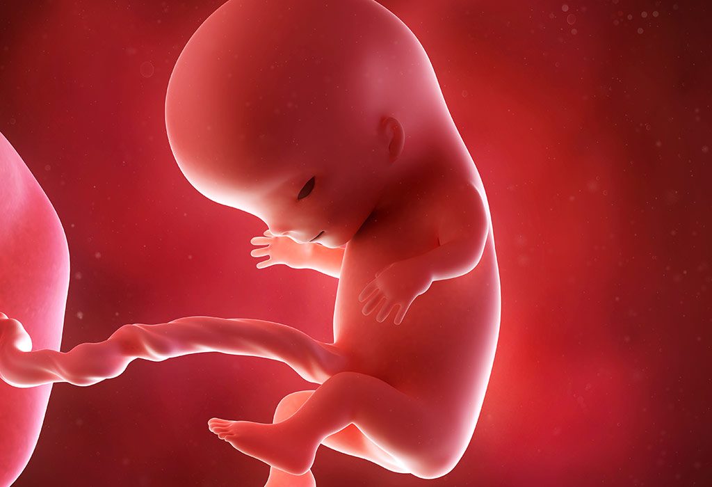
11 Weeks Pregnant – Symptoms, Baby Development, and Care
At eleven week pregnant, you’re entering the final stages of the first trimester, and your body is undergoing significant changes as it adjusts to pregnancy. 11 weeks pregnant is an exciting time as your baby has grown into a fetus from an embryo and formed all the workings of a human being; your body has seen changes such as a rounder structure, firmer uterus, rounder and more sensitive breasts. While symptoms like nausea and fatigue may persist, many women start to feel better as their hormones stabilise. Let’s progress to the next week of your pregnancy, the 11th week and explore the new exciting possibilities in your pregnancy.
Your Baby’s Growth During Pregnancy – Week 11
Pregnancy at 11 weeks is an exciting as well as wonderful time. At 11 weeks, the embryo is now considered a fetus and is rapidly developing. The 11th week is as fateful as the 9th and 10th weeks, as your baby looks more like a functional human being. For one, the facial bones of your baby are formed and hardened. The skin, however, is still transparent. Your baby is already starting to wiggle, but it’s not enough for you to realise yet.
You should be able to count 20 fingers and toes by now, seeing that your baby has developed arms and legs that have separated. More importantly, your baby has learnt how to swallow and is of course surrounded by amniotic fluid. The hair follicles are also in place, as are nipples, tongue and a nose with nasal passages (2) (3).
11 Weeks in Pregnancy Is How Many Months?
At 11 weeks pregnant, you are in the 3rd month of pregnancy, which marks the end of the first trimester. Pregnancy is typically divided into three trimesters: the first trimester (weeks 1 to 12), the second trimester (weeks 13 to 26), and the third trimester (weeks 27 to 40). Since you’re at 11 weeks, you are nearing the end of the first trimester and about to enter the second trimester.
What Is the Baby Size?
The 11-week fetus size is as big as a lemon or a fig. So, you can expect to see an 11-week pregnant baby size of 1-2 inches long. Since your baby’s body is also strengthening, the torso has also lengthened. Your baby weighs around 7 or 8 grams at week 11. The most interesting part is that your baby’s head takes up most of the formation, with a 1:1 proportion with the body.
Common Body Changes
It’s safe to say that pregnancy changes life, and your body will agree that it will never be the same again. As body changes during pregnancy are inevitable, let’s see what week 11 of your pregnancy holds (1):
Though still hard to tell, your pregnant body should have started showing the tell-tale signs of a bigger bump than during week 10.
For ladies who like curves, your body during pregnancy will certainly please you, with rounder features.
For the lucky ones, you may have the pregnancy glow that is usually much coveted. However, some women may end up with some pimples or rashes due to the increased production of hormones.
You have most certainly gained weight and also appear bloated.
Symptoms of 11th Week Pregnancy
Whether you are clearly showing a baby bump or not, and whether you have had any other changes in your body or not, you will most certainly show the following symptoms during the 11th week of your pregnancy. Examine the list below to compare the 11 week pregnant symptoms with what you are experiencing (4).
Leg cramps: This is probably one of the biggest events of the 11th week of your pregnancy. These cramps will manifest during the day, especially if you continue with your usual working schedule. However, be prepared for some interruptions while you sleep, as painfully tight leg muscles can disturb your sleep.
Linea Nigra: This symptom is exclusive to the 11th week of pregnancy. This symptom will manifest as a dark line right down the centre of your belly. To put you at ease, this is common and not permanent. Hormonal changes generally contribute to linea nigra.
Apart from the fresh symptoms stated above, the following symptoms from the last few weeks of your pregnancy will continue to manifest.
Fatigue: No surprises there. As your baby grows, your body will also feel heavier and seek rest.
Gas and Bloating: As you have seen in the past couple of weeks, your tummy will certainly continue to give you trouble as progesterone tends to slow down digestion.
Morning Sickness and Nausea: A few lucky ones might have this under control, but for most, continued trouble from both till you finish the first trimester.
Vaginal Discharge: While your sex life is bound to get better if you decide to indulge, invest in some pantyliners as the discharge will continue to increase as the pregnancy progresses.
Mood Swings: Your mood may change faster than traffic lights, and it’s best if you stay away from crowds as stress makes it worse.
Belly at 11 Weeks of Pregnancy
Most ladies may still be able to dismiss your pregnancy as gas or bloating. Nevertheless, your baby bump must already start to show, at least a little bit. For women who are pregnant for the first time, your eleven weeks pregnant belly may not appear to be different. For women who have been pregnant before, your tummy will likely hint at pregnancy.
Of course, it’s the opposite if you are on the slimmer or heavier side of the weighing scales. For heavier women, it might not seem different at all. For thin women, you might show a bit, especially if you have gained some weight.
11 Weeks Ultrasound
An eleven-week ultrasound will show how your baby is doing inside your belly. Your little baby’s movements will be graceful and fluid. Besides that, your baby’s skin is steadily going to become opaque as opposed to transparent. However, in week 11, it will remain transparent (3).
What to Eat?
One of the most important factors in this period is the concern about 11th-week pregnancy food. Read on to find what you must put on your plate in this period (8).
Foods rich in calcium: Since your baby’s bones have started forming, it’s important that you up the intake of calcium. Milk and milk products should be on top of your list.
Proteins: Lean meat and fish with a smattering of red meat is advised. Proteins help in the formation of bones and body mass.
Pulses and beans: These contain easily digestible proteins and several vitamins.
Seeds, sesame and sunflower: These are rich in vitamins and nutritious oils
Nuts, almonds and cashew nuts: Packed with protein and vitamins, nuts are a great addition to your diet.
Spinach: Rich in iron and packed with flavour, spinach is a superfood you are sure to like.
Yoghurt: A delicious source of calcium, yoghurt is a probiotic which will ensure the balance of good bacteria in your gut.
Fruits and leafy vegetables: Rich in vitamins and minerals, fruits and leafy vegetables ensure all-round development.
Tips & Care
Pregnancy can be challenging and, quite frankly, will take a toll on you. Nevertheless, keeping yourself and your baby safe and healthy should be of your utmost concern (2) (5).
Dos
Drink plenty of water to remain hydrated as well as curb leg cramps.
Plan a small vacation or a babymoon to relax yourself.
Be cheerful and read books on parenting to prepare yourself for parenthood.
Keep your doctor on speed dial so that they can always be contacted quickly.
Don’ts
Take any medication without consulting your doctor, as it could affect you or your baby’s health adversely.
Suppress your urine because frequent urination is a part of week 11 of pregnancy.
Indulge too much in junk food as your digestive process is already slowed down due to progesterone.
What You Need to Shop For
Being pregnant does not mean you cannot be stylish. Invest in good bras that stretch. You can also buy nursing bras at this stage that will come in handy later. Buy books on parenting that will give you information. Invest in maternity clothes and stretchy pants, preferably in cotton. Maternity kurtis are a good option as they’re super comfortable and offer ample space for the growing belly. For maximum comfort, while sleeping at night, Bella Mama’s maternity night suits would be perfect during pregnancy and even after pregnancy as they come with two side zippers for easy nursing. Stock up on the vitamins and health supplements that your doctor advises. Invest in comfortable shoes that will help with leg cramps and swelling. Do buy good oral care products that can help keep oral problems at bay.
FAQs
1. Is it safe to exercise at 11 weeks pregnant?
In most cases, exercising at 11 weeks pregnant is safe as long as your doctor approves. Low-impact activities like walking, swimming, or prenatal yoga are great options. Avoid high-risk activities and listen to your body to ensure you’re not overexerting yourself (7).
2. How much weight should I gain by 11 weeks pregnant?
Weight gain varies by individual, but most women gain between 1 and 5 pounds during the first trimester. Your doctor can guide you on what is healthy for your situation. Weight gain should be gradual, and it’s important to focus on eating nutritious foods.
3. Is it normal to have spotting or light bleeding at 11 weeks pregnant?
Light spotting or bleeding can occur early in pregnancy, but it’s important to mention it to your healthcare provider to rule out any complications. If the bleeding is heavier or accompanied by cramping or pain, seek medical advice immediately to ensure everything is okay with your pregnancy (5).
4. Can I travel during my 11th week of pregnancy?
Travelling is safe during the early stages of pregnancy, especially if your pregnancy is uncomplicated. However, taking frequent breaks is essential to stretch your legs, stay hydrated, and avoid heavy lifting (6).
While your baby bump is barely discernible, there are a lot of bodily changes you will have to deal with because of the hormones being produced by your body. The good news is that a healthy diet and comfortable clothes can go a long way in bringing comfort.
Previous Week: 10 Weeks Pregnant
Next Week: 12 Weeks Pregnant
Read more

34 Weeks Pregnant: Symptoms, Baby Development, & Care Tips
At 34 weeks pregnant, your baby is continuing to grow and mature while your body prepares for the final weeks before birth. You may notice more fatigue, back pain, heartburn, swelling, frequent urination, or difficulty sleeping as your belly gets bigger. Your baby is also becoming more active, although the way those movements feel may change as space inside the uterus becomes limited. This stage is an important time to keep up with prenatal appointments, pay attention to your baby’s usual movement pattern, and watch for signs that need medical attention. In this article, we’ll cover what’s happening with your baby at 34 weeks, common pregnancy symptoms, important care tips, and when to contact your doctor.
Your Baby’s Growth During Pregnancy – Week 34
Your 34th week of pregnancy is going to show well-established sex markers, with boys and girls easily differentiable. (Please note that even if your doctor knows the sex of the baby, you won’t be told, as sex determination is illegal in India.) Your baby’s fat has now fully filled out the wrinkly skin, making it smooth and robust. The immune system and lungs get stronger and more independent of your body (1). As the foetus’s brain connections now number in the trillions, it is even possible for it to dream occasionally!
Your uterus is now at near-maximum capacity, and any shift in position will be easily felt by you. In fact, your baby might even be able to perceive your warm touch through your abdomen. Preterm delivery around this point is completely safe, excluding a short stint in the hospital, and you will be ready to take your little one home in no time. Don’t worry if you don’t experience labour around the due date, as that only happens in five per cent of all cases.
What Is the Baby’s Size at 34 Weeks?
At 34 weeks pregnant, the baby’s size is almost 45 cm from top to bottom – around the size of a large pumpkin. At slightly over 2 kg, your baby is still going to grow some more (2). Its skeleton is hard by this time, except for the skull bones, which remain soft to allow passage through the birth canal.
Common Body Changes
Week 34 will bring its own challenges to the table. This week you will experience another host of body changes during pregnancy.
1. Haemorrhoids
Haemorrhoids are caused due to factors such as constipation and hormonal activity during pregnancy (3). Much like last week, you stand the risk of developing painful swellings in the anal or perineal area. Medications and Kegel exercises are options for some relief.
2. Discharge From the Vagina
Another common symptom is mucous secreted by your vagina in preparation for birth. This is caused by the influx of oestrogen, which promotes blood circulation around the vagina (4).
3. Swollen Feet
Your feet and ankles might continue to suffer from oedema due to all the fluid collecting in them. Opt for comfortable footwear that has soft padding to ease your swollen feet (5).
4. Stretch Marks
If you are at your 34th week of pregnancy, these cannot be avoided! However, their extent can be reduced by controlling your weight gain under medical supervision.
5. Discharge From Breasts
Your body is making colostrum for when your baby is born, and your breasts have decided to leak it early (6). Wear absorbent pads inside your bra to avoid embarrassing situations.
34 Week Symptoms of Pregnancy
The following are the 34-week symptoms of pregnancy (5):
1. Constipation
The expanded uterus now takes up nearly all your abdominal space, forcing your bowels into a corner. This will tend to make your bowel movements rather irregular. Consuming enough fibre and water should ease the discomfort.
2. Bloating
Gas is extremely common during pregnancy due to the slowing down of digestion, with anxiety worsening the situation. Try a few breathing exercises to ease the stress.
3. Cramps
Carrying around all that weight and lack of sleep, band together to create painful leg cramps.
4. Difficulty Breathing
A frequent symptom in late-stage pregnancy, difficulty breathing is one of the 34 weeks pregnant symptoms not to ignore if it becomes sudden, severe, or occurs even while resting. It can happen because the growing uterus puts pressure on the diaphragm, making it harder to take a deep breath. Try to slow down and avoid strenuous activities when you feel breathless.
5. Staying Up All Night
Anxiety, cramps, bloating, and constipation are going to keep you up all night. If you just can’t fall asleep, try watching television or reading a book.
6. Backache
As your belly grows and your centre of gravity shifts, the extra weight can put more strain on your lower back. Gentle stretching, good posture, and using a pregnancy support pillow may help ease the discomfort.
7. Swollen Feet and Ankles
Mild swelling in the feet and ankles is common during the later stages of pregnancy because your body retains more fluid and the growing uterus can affect blood circulation. Resting with your feet elevated and avoiding standing for long periods may help.
Belly at 34 Weeks of Pregnancy
In your 34th week, you can expect your abdominal region to stretch to around 45 cm from the top of the uterus to the lower pelvic region. An interesting thing to note is that the volume of the amniotic fluid is much higher at this point than at any other point during your pregnancy. Your uterus is now up to 1,000 times bigger than before it had a baby in it.
34 Weeks How Many Months Pregnant?
At thirty four weeks pregnant, you are about 7 months and 3 weeks pregnant, which means you’re in your 8th month of pregnancy and approaching the third trimester’s final stretch.
Ultrasound at 34 Weeks Pregnant
Going for ultrasound scans every alternate week is standard procedure at this point. Your doctor might even prescribe a non-stress test or a biophysical profile, which can tell if there’s something wrong with the baby’s breathing or heart. The scan can show your baby’s movements in real-time, such as breathing or moving around the uterus. If you want, you can see the intimate details of your baby’s face and body with a 3D ultrasound.
What to Eat When You Are 34 Weeks Pregnant?
Eating a balanced diet is crucial for your and your baby’s health. Consume several portions of fresh fruits, vegetables, whole grains, and dairy a day. Eat frequent but small meals so you can avoid heartburn. Some of the dietary changes to make in your 34th week pregnancy food are:
Vitamins D, C, and K are essential in bone and teeth development, as well as boosting immunity. Vitamin D can be consumed via supplements, but exposing your body to sunlight for 10-20 minutes a day also does the job. Vitamin C can be found in leafy greens, lean meats, and citrus fruits. Vitamin K can be found in foods like cauliflower, cabbage, beans, pomegranate, spinach, and soybean (7).
It is certainly time to have omega-3 fatty acids to have your baby’s intellectual capacity reach its full potential. Fish oil is available to suit any interest.
Along with following the 34-week pregnancy diet, drink 3 litres of water a day, minimum, to stay fit for your delivery.
Tips & Care
Below is a set of activities that you should and should not do while you’re thirty-four weeks pregnant:
Dos
Perform some exercises to ensure your baby is in the ideal position for delivery, that is head downwards with his face towards your spine. Some exercises use a chair or birthing ball, so you can exercise by keeping your pelvis leaning forward.
It might help to visit the hospital where you are thinking of giving birth. Observe the labour and birthing wards of the hospital, and complete any paperwork you might have to.
Don’ts
Dieting is not fun, and while you’re in the third trimester, it’s outright dangerous. Your baby and you both need considerably more food than usual, so cutting them out will cause complications. Instead, ignore unhealthy foods such as alcohol and spicy food.
Don’t forget to perform Kegel exercises, and don’t be embarrassed about them! Kegel exercises make the pelvic floor stronger, and they can ease pain caused by cramps and hemorrhoids. In addition, the exercise improves the chances of a smooth delivery. You can also perform the pelvic tilt, which can help ease any backache.
What You Need to Buy
Make sure your baby has a nice, warm home to be welcomed into. Maternity clothes from Bella Mama are always first on the purchase list, and there are several stylish options available. If you prefer to wear Indian ethnic clothing on most days, maternity kurtis are a must-have for you as they provide ample room for the growing belly. Maternity tops are another favourite as they are available in lovely silhouettes, prints and colours.
FAQs
1. What are the signs of preterm labour at 34 weeks?
At 34 weeks, signs of preterm labour may include consistent contractions every 10 minutes or more, lower back pain, pelvic pressure, and changes in vaginal discharge. It’s important to contact your healthcare provider immediately if you experience these symptoms, as timely intervention can be crucial.
2. How can I manage increased fatigue at this stage?
Increased fatigue at 34 weeks can be due to the physical demands of carrying extra weight and preparing for labour. To manage this fatigue, prioritise short naps throughout the day, consider gentle prenatal yoga or stretching, and ensure you’re staying well-hydrated and nourished.
3. Is it normal to experience heightened emotions or mood swings?
Yes, heightened emotions or mood swings can occur during the final weeks of pregnancy due to hormonal changes and anxiety about childbirth. Engaging in relaxation techniques, such as mindfulness or deep- breathing exercises, can help manage these emotions.
This was all about pregnancy at 34 weeks. Keep sufficient stocks of diapers, bottles, nursing bras, and baby clothes. Buying a little pram will also make it easier for you to get your baby to your home or neighbourhood. As the time draws closer towards your final day, do your best to stay calm by talking to your friends and family. You’re almost to the finish line, so just keep doing what you’re doing, and you’re as good as gold!
Previous Week: 33 Weeks Pregnant
Next Week: 35 Weeks Pregnant
Read more

16 Weeks Pregnant: Symptoms, Baby Development, Diet & More
At 16 weeks pregnant, your baby is around 11.6 cm long, about the size of an avocado, and is becoming increasingly active as their nervous system develops. You may also notice your own body changing, with symptoms such as constipation, bloating, headaches, heartburn, or round ligament pain becoming more noticeable. Some women may even begin to feel the first gentle flutters of movement, although it is completely normal not to feel them yet; most people notice movement between 16 and 24 weeks. In this article, we’ll cover what’s happening to your baby and body at 16 weeks, common symptoms, healthy diet choices, lifestyle tips, prenatal care, and when to contact your doctor.
Growth of the Baby at 16 Weeks of Pregnancy
This week is the real deal, as your baby is likely to have fully formed genitals and eyes that are still fused but sensitive to light. Your baby may also start yawning inside your tummy now. The tiny muscles and backbone have gained strength, which means that your baby can now straighten their head and neck further than in the last few weeks. Your baby’s facial features have matured, thereby enabling him to frown or squint (1). However, your baby’s skin will still be transparent (2).
Size of the Baby
This is probably the most exciting part, as your tummy is now home to a 4-5-inch foetus that weighs around 100 grams (3). The other exciting part of your baby’s size is that your doctor will now know if your baby is a girl or a boy as the genitals are fully formed. Let’s just say that your avocado-sized baby is as good as a fully functional human being (4). Keep in mind that at 16 weeks pregnant, the baby’s size may also vary depending on different medical factors.
Common Bodily Changes
Since your baby’s body and weight have already changed at the onset of 16 weeks, it is hardly a surprise that you will face certain changes in your body during pregnancy at the end of this week. For starters, you will be curvier than before and may have to wear bigger clothes.
You will notice that you have fewer mood swings and a beautiful glow on your face, and you will feel less nauseated (5). Your biggest worry at this point will soon be stretch marks besides the well-being of your baby.
Symptoms of Pregnancy at Week 16
By this time, you are likely to get adjusted to the usual symptoms of pregnancy. It is, of course, safe to say that some symptoms are likely to continue until the end of pregnancy, while new ones will develop as each week progresses. Here are some common sixteen weeks pregnancy symptoms:
You may experience backache now owing to the weight of the foetus (6).
Your breasts will certainly be bigger and will continue to be sensitive.
Your eyes will be more sensitive owing to the changing hormones.
You may end up being constipated more often (4).
You may also be forgetful owing to the hormonal changes that your body is constantly undergoing.
You might also get varicose veins or cramps in your legs (1).
Nasal congestion or a stuffy nose can also occur because hormonal changes increase blood flow to the nasal passages.
Belly at 16 Weeks of Pregnancy
At times, you might struggle to catch your breath. This is because your baby is growing, gaining weight and generally feels free to move around your belly. More importantly, there will be quite a bit of activity inside your belly as your baby might have started kicking or simply moving around, and you might want to be careful because muscle twitches and gas are still common in the 16th week of pregnancy.
16 Weeks Is How Many Months in Pregnancy?
16 weeks gestation is four months pregnant. It is important to note that doctors track pregnancy progress in weeks, not months.
Ultrasound Scan at 16 Weeks of Pregnancy
The ultrasound at the 16th week will show the maturation of organs. Your baby’s taste buds will form, and so will his eyebrows, hair, and eyelashes. Since your baby can now identify voices and sounds, it indicates that the tiny bones of the ears, as well as the tympanic membrane, will have developed.
In addition to the 16-week sonogram, your urine might also be tested during this week to check for diabetes and other diseases. Besides that, the usual signs of your baby’s growth, such as heartbeat, maturing organs, movement and sensitivity towards light, can all be charted.
What to Eat
Apart from the well-being of your child, the other thing you will mostly worry about at the 16th week of your pregnancy is food. Listed below is a suggestion of what you should be eating.
A bite of chocolate may do you good, but as a regular practice, continue eating fruits and yoghurt, which aid digestion and provide necessary nutrients.
String cheese is an excellent choice for those craving salty foods, as it is rich in calcium and proteins.
Include a range of fresh fruits and vegetables in your diet to make sure your baby gets the nutrients essential for growth (7).
Make sure you consult a doctor and take proper vitamin supplements. These ensure that you get the nutrition that food can alone not suffice.
Lean meat, little red meat, fish, eggs, and pulses are all good choices to include in your diet as they contain easily digestible proteins.
Make sure you include iron, vitamin C, and calcium in your diet to supplement it.
Care Tips
This is the time when you should start taking extra care of yourself. Eating properly will benefit your weight gain, as it is important to have the strength to bear the weight of your baby. Make sure you do not perform any task in excess as this may have a negative effect on your unborn baby.
Dos
Exercise mildly or walk, at least for 30 minutes each day, to relieve your backache or leg cramps (8).
Remember you aren’t eating for two. You are simply providing nutrition to yourself and your baby. So, go for mindful eating and do not overeat.
Get plenty of rest and adequate sleep.
Stay hydrated without increasing soft drink consumption.
Think positive and stay cheerful, as that will go a long way to improving mental health.
Don’ts
Drink too much caffeine as it can have a negative impact on your baby’s overall health.
Indulge in heavy workouts, as they may leave you exhausted.
Starve yourself or your foetus.
Stay in an unhygienic state.
Ignore your oral health.
Stress yourself out.
Shy away from asking for help as and when required.
What You Need to Shop For
Pregnancy doesn’t need to be boring; flaunt your baby bump in style. If you prefer stylish, shop for bright maternity dresses that make you feel comfortable and good. Invest in good shoes, preferably with soft padding that will relieve the muscles of your feet. Comfortable tops and button-up maternity kurtis look great and are very convenient, too. Bella Mama has a lovely range of maternity night suits and nighties that are ideal as comfortable sleepwear. Get a good anti-stretch mark cream or oil because your skin will keep expanding. Do buy some lotions and creams because your skin will feel dry. Don’t forget to invest in a practical handbag that can stock up your essentials. Also, purchase eye drops that provide a cooling effect that can help relax your eye muscles. Oral care products are important,t too. Finally, don’t forget to purchase good parenting books.
When to Consult the Doctor?
During the second trimester, you may have prenatal appointments with your doctor every 3-4 weeks or differently depending on your progress. However, if you feel something unusual or any sudden discomfort, it is advised that you don’t wait for your scheduled appointment and consult a doctor as soon as possible to get your concerns checked.
At 16 weeks of pregnancy, you should immediately consult a doctor if you experience:
Vaginal bleeding (should always be informed to the doctor) (9)
Severe cramping
Fever
Dizziness or fainting
Changes in vaginal discharge
Foul-smelling discharge
Severe vomiting
FAQs
1. Where is your baby positioned at 16 weeks of pregnancy?
By 20 weeks, the uterus slowly reaches the belly button. In most pregnant women, the 16-week fetus is usually below the belly button.
2. Can you feel the baby when 16 weeks pregnant?
Most pregnant women feel the movements of their babies for the first time between 16 and 20 weeks of pregnancy (10). This phenomenon is called quickening (11). The movements may feel like a little fluttering, tiny pulses, or bubbles in your tummy. However, if you don’t feel your little one’s movements between 16 and 20 weeks, please remember that it is normal.
3. Why do I frequently feel so constipated?
Constipation in pregnancy is a common pregnancy symptom throughout the pregnancy, which is caused by a combination of factors, like prenatal vitamins or low activity levels (12). Although not dangerous, it could lead to discomfort and other issues, like haemorrhoids and anal fissures. Contact your doctor for a suitable remedy.
4. How can I prevent pregnancy stretch marks?
Honestly, there’s not much you can do to avoid pregnancy stretch marks, as it is genetic. But the best you can do to stay away from them is to stay hydrated by consuming water and water-rich foods and moisturising your skin regularly.
A thorough idea of what to expect during pregnancy can keep you ready to overcome the challenges that you may likely face during this period. Don’t stress; enjoy each and every moment, and have a great pregnancy!
Previous Week: 15 Weeks Pregnant
Next Week: 17 Weeks Pregnant
Read more
28 Weeks Pregnant – Symptoms, Baby Size, Belly & Care
At 28 weeks gestation, you are entering the third and final trimester of your pregnancy, which is expected to last until week 40, when you are most likely to give birth. Isn’t that an amazing feeling? The third trimester typically lasts from the 7th month to the 9th month of pregnancy, and there is a high chance that labour pains may start a couple of weeks before the due date. At this crucial juncture of the 28th week of pregnancy, your doctor may ask you to visit at least once every 2 weeks, depending on your pregnancy progress. In this article, we’ll discuss everything you need to know about the 28th week of pregnancy, including key symptoms, your baby’s growth, dietary tips, and self-care recommendations.
Your Baby’s Growth During Pregnancy – Week 28
By week 28, your baby can blink her eyes, and her eyesight is developing. She is able to see the faint light entering your womb. Her little brain is also developing billions of neurons and can perform tasks such as blinking, dreaming, and controlling her body’s temperature (5).
The biggest transformation in the third trimester is the development of your baby’s bones from cartilage, which happens in the 7th and 8th months. Make sure that you eat a lot of calcium-rich foods, as your baby requires all the calcium for bone development.
Your baby will also develop all her five senses during the third trimester. This would include the development of her touch receptors (around week 29 or 30), along with other sensory signals like light perception, tasting what you eat, and listening to your voice, all of which happen by week 31.
28 Weeks in Pregnancy Is How Many Months?
At twenty-eight weeks pregnant, you are in the seventh month of pregnancy. With only about 12 weeks to go, this is an exciting and crucial time as you prepare for the arrival of your baby (4).
What Is the Baby’s Size?
When you are 28 weeks pregnant, the baby’s size is around the size of a large eggplant, and it will weigh around 2.25 pounds (approximately 1.02 kg). The baby is now about 14.25 to 14.80 inches long (1).
The third trimester is also the fastest period of growth for babies, as they grow from 16 inches to around 19-22 inches by the 40th week of pregnancy. Along with height, the weight of your baby also increases at 28 weeks gestation. From about 1 kilogram at the beginning of the 28th week to between 2 and 4 kilograms by the 40th week, your tiny human grows bigger by the day!
Measurement
Progress
Length (crown to heel)
About 14.80 inches
Weight
About 2.22 pounds (1 kg)
Skills
Can hiccup, suck, kick, blink, roll, and grasp
Common Body Changes
With all the baby development happening inside your 28 weeks pregnant belly, you are going to experience more body changes during pregnancy in this final trimester.
Common body changes that occur during this pregnancy period can include:
The appearance of stretch marks on your baby belly: These are tiny tear marks that appear on your belly skin and are caused by the skin being stretched during the final phase of your pregnancy. This could also be genetic in nature, so if your mother had them during her pregnancy, you are also likely to develop stretch marks.
Lack of sleep at night: This is a common occurrence in this trimester, and is caused by factors like your baby’s kicking, the large belly, frequent trips to the bathroom, and several other reasons.
Swelling of your feet: Also known as oedema, this is caused by water retention as well as the increase in weight.
Other common body changes during the third trimester can include leaking breasts and bulging of veins in your lower body (5).
Symptoms of Pregnancy at Week 28
Other than the body changes, 28 weeks pregnant woman may experience a few mild or severe symptoms during the third trimester (1) (2):
Pain in the abdomen: You may experience cramping or sharp pain in your lower abdomen as it stretches to accommodate your growing baby bump.
Fatigue: In the third trimester, your pregnancy demands a lot from your body, resulting in regular fatigue. You must eat well and be active to avoid excessive fatigue.
Backache: The growing belly will put a lot of pressure on your backbone, thus causing backache. It is essential to keep both your feet up to reduce the stress on your back.
Restless leg syndrome: Some pregnant women may experience a creepy sensation in their lower limbs, resulting in an urge to keep moving their legs, even when sleeping or relaxing. Stretching or massaging your legs can help relieve restless leg syndrome, or you can consult your doctor for iron supplements.
Preeclampsia: This occurs in women with high blood pressure after 20 weeks of pregnancy. Common symptoms of preeclampsia include significant swelling on your face or puffiness around your eyes, rapid weight gain, persistent headaches, nausea, or vomiting. Severe preeclampsia can lead to life-threatening problems; hence, it is best to consult your doctor if you have any of these symptoms.
Leaky breasts: By now, your body is preparing food for your baby; however, you may have to deal with some milk leaking out of your breasts.
Shortness of breath: As your baby continues to grow and your belly expands, it puts immense pressure on your lungs, because of which you may feel you need to catch your breath from time to time.
Other common symptoms that you may experience include dizziness, bloating or gas problems, stuffy nose, and bleeding gums.
Belly at 28 Weeks of Pregnancy
At 28 weeks, your baby should get in the right position (head downwards) inside your belly in preparation for labour. The fundal height of your belly, which is the distance from the pubic bone to the top of your uterus, should measure around 26 to 30 centimetres. If you are expecting twins, the measurement of your fundal height may not be possible.
You must now start keeping tabs on whether your baby is moving consistently through the day. Check how long your baby takes to reach ten foetal movements in a day. Ideally, it should be less than 2 hours.
28 Weeks Ultrasound
After 28 weeks of pregnancy, your baby is developing more fat, so her skin is starting to get smoother. Your baby’s lungs are mature enough to begin the process of breathing.
Unless there are any pregnancy-related complications, a 28-week ultrasound would not be recommended by any doctor. You would probably start seeing your doctor more often, every 2 weeks or twice a month.
Tests and Checkups Around 28 Weeks
Around 28 weeks of pregnancy, antenatal care becomes increasingly important as you enter the third trimester. Your doctor will monitor your health and your baby’s growth, and recommend tests depending on your medical history, previous test results, and pregnancy complications, to check for conditions that can affect pregnancy and delivery.
Common checks around this stage may include (9):
Blood pressure and weight: These are routinely monitored during antenatal visits and can help identify problems such as high blood pressure and preeclampsia.
Blood glucose screening: Screening for gestational diabetes is commonly performed between 24 and 28 weeks of pregnancy. A glucose screening test may be followed by a diagnostic test if the initial result is elevated (8).
Anaemia testing: Your doctor may check haemoglobin levels, particularly if you have risk factors or symptoms suggesting anaemia. WHO includes haemoglobin testing among routine antenatal blood investigations (9).
Rh status and anti-D care: If you are Rh-negative, your doctor may discuss anti-D immunoglobulin around 28 weeks to reduce the risk of Rh sensitisation.
Baby’s growth and fundal height: After 24 weeks, fetal growth and fundal height are generally monitored during antenatal contacts.
Fetal movements: Your doctor may discuss monitoring your baby’s movements as part of third-trimester care.
What to Eat?
As your baby experiences the fastest growth during this trimester, your 28th-week pregnancy food will help supplement her growth. However, due to the size of your baby’s belly and regular nausea, you may not feel like eating a lot. So, instead of quantity, opt for quality foods that are rich in essential nutrients. Instead of large meals, opt for six or more mini-meals or snacks throughout the day.
Doctors also recommend eating fish during the third trimester, as fish provides a lot of health benefits to your baby. Avoid fish like king mackerel and swordfish, as they contain high quantities of mercury.
Tips & Care
As you approach delivering your baby, here are essential tips and do’s and don’ts (6).
Dos
Listed below are some pointers that you should keep in mind during the third trimester of your pregnancy.
Ensure that you schedule and go for your 28-week prenatal appointment.
Keep a regular count of the foetal kicks.
Make sure you get lots of sleep.
Maintain your daily exercise schedule.
Don’ts
Listed below are some things you should not do at any point in your pregnancy.
Avoid smoking as it can lead to lower baby weight and respiratory problems.
Don’t consume alcohol, as it can significantly impact your baby’s overall development.
Do not eat raw or undercooked meat or eggs.
Avoid deli meat such as hot dogs and sausages.
Avoid sitting in a sauna or a hot tub.
What You Need to Shop For?
The rapid growth of your baby belly can create a lot of discomfort in the abdominal area, and this puts pressure on your backbone. A maternity bra is an absolute must-have item during pregnancy. As the rib cage expands because of the growing baby, you’ll need to find a good, comfortable wireless bra. Also, check out the maternity nighties from Bella Mama. These are knee-length nighties with concealed zippers that provide good access for nursing. You can shop for a maternity belt if you are experiencing severe pain in the hip and pelvic area. A belly wrap would also be an excellent buy to reduce the strain on your back. Knee-high maternity compression socks might also be a worthy purchase to provide you relief from your leg muscle ache and varicose veins.
Questions to Ask Your Doctor
Your 28-week appointment is a useful time to discuss your baby’s growth, upcoming tests, nutrition, symptoms, and preparations for the final trimester. These questions will help get an overall picture of your current pregnancy status:
Is my baby’s growth and development appropriate for 28 weeks?
How should I monitor my baby’s movements, and what changes should prompt me to contact you?
What foods, nutrients, or supplements should I focus on during the third trimester?
How much weight should I expect to gain during the remainder of my pregnancy?
What warning signs should make me contact you or seek urgent medical care?
What should I start preparing for as I approach the third trimester and delivery?
When to Consult a Doctor?
You should consult your doctor immediately at 28 weeks if you notice reduced fetal movement, signs of preterm labour (regular contractions, fluid leakage, or bleeding), or symptoms of preeclampsia like severe swelling, persistent headaches, vision changes, or sudden weight gain. Other red flags include severe abdominal pain, unusual vaginal discharge, shortness of breath, chest pain, high fever, or signs of gestational diabetes such as excessive thirst and fatigue (5).
FAQs
1. Can I exercise at 28 weeks?
Yes, light to moderate exercises like walking, swimming, or prenatal yoga are beneficial for maintaining fitness, reducing stress, and preparing for labour. However, avoid high-impact or risky activities (3).
2. Should I start preparing for labour and delivery at 28 weeks?
It’s a great time to start planning. Consider taking a childbirth class to learn about labour, delivery, and newborn care. Discuss your birth plan with your doctor or midwife, and start packing a hospital bag with essentials like clothes, toiletries, and important documents.
3. Should I get vaccinated during the third trimester?
Certain vaccinations, such as the Tdap vaccine, are recommended during the third trimester to protect your baby from illnesses like whooping cough. Discuss your vaccination schedule with your doctor to ensure you and your baby are protected (7).
In the third trimester, you may be excused for feeling just a little tired of the discomforts that have been plaguing you for some time now. The good news is that delivery is almost around the corner, and soon the struggle of the last few months will be rewarded with the miracle of birth.
Previous Week: 27 Weeks Pregnant
Next Week: 29 Weeks Pregnant
Read more
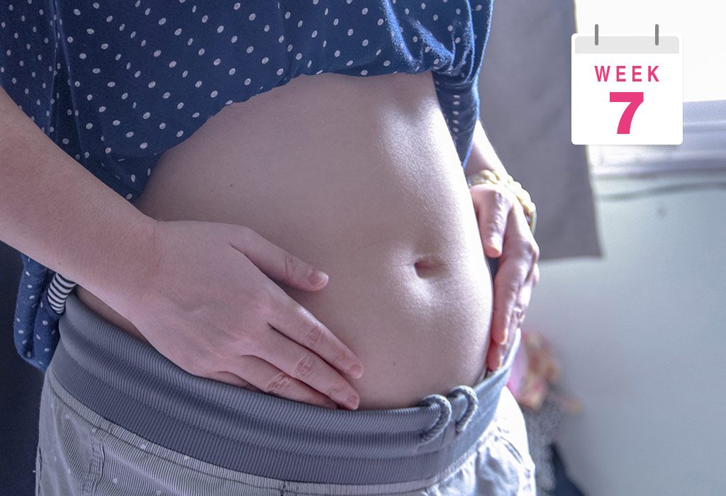
7 Weeks Pregnant – Symptoms, Baby Growth and Care Tips
In week 7 of pregnancy, you can expect fast and exciting developments. Being 7 weeks pregnant, you must be experiencing the symptoms of pregnancy, such as morning sickness, nausea, and more, but you don’t quite look pregnant yet. Your baby is growing rapidly inside your 7 weeks pregnant body. This is the period of major developmental changes for you and your baby. Your body will start preparing itself to nurture the growing foetus. This is also when you have to be very careful about what you eat and avoid, and how you take care of yourself. If you have any doubts, we recommend reading this article to learn how your little one is developing and what changes you can expect in your body during this week.
Your Baby’s Growth During Pregnancy – Week 7
Your baby’s legs and arms will begin to emerge by this time. The umbilical cord has formed and your baby will have a small tail, which is more like an extension of the tailbone (1). However, this will disappear as the pregnancy progresses. The foetus has almost doubled in size in the past week. Your baby will experience rapid developmental changes. Both the hemispheres of the brain will start forming now. The tiny liver will begin to function by forming red blood cells till the bone marrow is formed. The eyelids, limb buds, and digestive organs like the pancreas and appendix start to form, too (2). At this stage, your baby may technically be called an embryo.
What Is the Size of the Baby?
Your baby is approximately the size of blueberry or a grape by this time. The 7 weeks pregnant baby size is almost an inch according to the American Pregnancy Association (3). There has been tremendous development from the time of conception as your baby has grown a lot since then and will continue to grow and develop as they should. At this time, the growth is mostly concentrated in the generation of brain cells.
Common Bodily Changes
You may not look pregnant, but several changes are happening inside your body. You can expect the following changes at around 7 weeks of your pregnancy (4).
Nausea and food aversions: You may experience nausea and food aversions by this time. Some odours or smells may trigger nausea. On the other hand, you may also find yourself craving for foods such as pickles.
Excess formation of saliva: You may notice the formation of excess saliva in your mouth. Try chewing sugarless gum to avoid the pooling of excess saliva in your mouth.
Changes in the breasts: Your breast size may increase this week. This is due to the increase in blood flow and fat build up around your breast.
More frequent bathroom visits: There is an increase in blood flow around your pelvic region because of the hCG hormone, resulting in frequent urination.
Skin breakouts or acne: You may also notice acne on your face. This is due to the hormonal changes happening in your body (5).
7 Weeks Pregnant Symptoms
Wondering what the seven weeks pregnancy symptoms are? Well, there may not be evident physical signs by 7 weeks of pregnancy, but you may notice the following symptoms this week (6):
You may start to feel sick in the mornings (nausea and vomiting), experience headaches and other pains, and notice fatigue and lethargy.
Your breasts may become tender, and the areoles will become darker than usual.
You may crave certain food items and develop an aversion to some.
You may not fit too well into your favourite pants or jeans. This is not due to weight gain but due to bloating.
You may also experience mild pelvic cramping. There might be occasional spotting, too.
One of the important developments in your body during this time is the development of your mucous plug. The mucous plug helps seal the cervix and protects the opening of the uterus till the birth of your baby.
You may find yourself battling with changing moods. This is due to the changing hormones in the body.
Belly at 7 Weeks of Pregnancy
Your belly at 7 weeks won’t give away that you are pregnant even though you might be experiencing a lot of changes in your body. So, you will have to wait for some time to be able to flaunt your baby bump. All that you feel is bloating around your belly by week 7 of your pregnancy. Most mothers-to-be start to show the baby bump by the middle of their second trimester. However, if you are carrying twins or more babies, then your belly may start to show earlier than the standard time.
Ultrasound at 7 Weeks of Pregnancy
By week 7, you may be ready for your first ultrasound scan, though many doctors may wait till 8 to 10 weeks of pregnancy for the scan. Your doctor will ask about your medical history and your doctor’s recommendation. An ultrasound will help the doctor take various measurements of your baby and determine the development of the foetus. Though your baby is too tiny for you to spot in the 7-week ultrasound scan, the embryo is generating almost one hundred brain cells per minute. The embryo is developing its heart, kidneys, arms and leg joints. If you are carrying twins, the scan will show two gestational sacs in the ultrasound scan. You will also be required to give urine and blood samples for various tests.
The blood samples of the mother carrying twins or multiple babies may show increased levels of hCG hormone, this increased levels of hormone may result in morning sickness.
What to Eat?
It is very important for you to eat a healthy and balanced diet in pregnancy. Your 7th-week pregnancy food should include the following (7):
A healthy mix of green, red, yellow and orange vegetables is a must include in pregnancy diet. Have 3 to 5 serving of vegetables in day. Spinach, broccoli, tomatoes, red peppers, pumpkin, carrots, yellow peppers and corn are some of the fresh vegetables you may include in your diet.
It is suggested to opt for fresh fruits over canned or preserved food as preserved food may contain harmful bacteria which can be harmful to you and the baby. You should take 3 to 4 servings of fresh fruits in a day.
Consume milk, cheese, yoghurt and other milk products to meet the calcium demands of your body. Around 3 servings a day is recommended.
You should include 2 to 3 serving of protein in the form of eggs, meat and poultry in your diet. Lentils, beans, seeds and nuts are excellent sources of protein for the vegetarian moms-to-be.
You should opt for whole grains instead of refined food products. Whole grains provide fibre to your body. It is suggested to have 3 serving of whole grain food items in a day.
Tips & Care
Morning sickness and nausea can take a serious toll on your health during this time. Here are some dos dos and don’ts that you should stick to for a healthy pregnancy:
Dos
Eat bland food and eat often as it is easier to digest.
Keep yourself hydrated.
Drink lemonade or eat watermelon to ease nausea.
Take adequate rest throughout the day.
Wear a good bra to ease breast soreness.
Take prescribed vitamin supplements.
Don’ts
Avoid spicy food.
Strictly skip alcohol and smoking.
Avoid skipping meals.
What You Need to Shop For?
Your shopping list at 7 weeks of pregnancy should include the following:
A Pregnancy Journal: To note down your feelings and emotions during pregnancy.
Good Lotions: You can buy good lotions or oils to moisturize your skin as it may become drier than before.
A Comfortable Brassier: Now that your breasts are tender and swollen, wearing a comfortable bra may be helpful.
Comfortable Bottom Wear: A must, might we say. It is recommended to opt for stretchable fabric as your size will constantly be changing in the coming months.
Good Pregnancy Books: You may buy good pregnancy books to know more about pregnancy.
FAQs
1. How many months is 7 weeks of pregnancy?
If you are 7 weeks pregnant, you are in the 2nd month of your pregnancy.
2. Is it safe to eat processed foods like instant noodles to ease cravings at this time during pregnancy?
Processed foods like instant noodles carry no nutritional value and contain low-quality ingredients, high sodium, food colours, preservatives, additives, and flavour enhancers. All these are harmful for a pregnant woman if she eats them frequently. Occasional eating is fine.
3. What is the usual “feeling” of being 7 weeks pregnant?
It is possible to probably feel nothing to some pregnancy symptoms, like bloating, cramps, mood swings, and fatigue, when you are seven weeks pregnant.
4. How does your tummy feel when you are 7 weeks pregnant?
If you’re dealing with morning sickness in the 7th week of pregnancy, it’s common for it to peak during the end of your first trimester. You might also have some gastrointestinal discomfort that makes your stomach feel unsettled.
5. Can pregnancy symptoms subside at 7 weeks of pregnancy?
Pregnancy symptoms often begin to ease after the first trimester. While you may notice a decrease in symptoms around this time or in the following weeks, every pregnancy is unique. Symptoms may linger for a shorter or longer duration, so they might not disappear entirely.
6. Can I get beauty treatments or dye my hair when 7 weeks pregnant?
Many beauty treatments involve the use of strong chemicals. Thus, certain beauty treatments, like laser procedures or skin peels, should be avoided. With hair dyes, if you are using brands that are ammonia-free and low-chemical options, then it is fine.
Your body will undergo a lot of changes in the 7th week of your pregnancy. But it’s just the beginning. You may feel nauseous, fatigued and lethargic. Things will get better in the second trimester, and morning sickness and nausea may return in the third trimester. But by staying positive and following a healthy lifestyle, you can have a healthy pregnancy. If you experience any discomfort, connect with a doctor immediately.
Previous Week: 6 Weeks Pregnant
Next Week: 8 Weeks Pregnant
Read more
5 Weeks Pregnant – Symptoms, Baby Size and Body Changes
At five weeks pregnant, things may not look very different on the outside, but plenty is happening inside your body. Your pregnancy hormones are rising, your baby is beginning some of the most important early stages of development, and you may start noticing changes such as tiredness, nausea, sore breasts, or mild cramping. It’s also perfectly normal if you don’t feel many changes yet. So, what exactly is happening during this early stage? Let’s look at your baby’s development, common symptoms, changes in your body, and the care tips that can help you have a healthy start to pregnancy.
Your Baby’s Growth During Pregnancy – Week 5
The embryo is growing fast inside you, and your baby now looks like a small tadpole with a primitive head and tail. The embryo now has three layers – the ectoderm, the mesoderm, and the endoderm. These three layers will later develop into various body parts and complete your baby’s features. The neural tube also starts to develop this week in the top layer or the ectoderm. Your baby’s brains, spinal cord, nerve, and backbone will all sprout out of the neural tube. His heart and circulatory system have started to form in the mesoderm. In fact, in the 5th week, your baby’s heart will start splitting into various chambers, beating and pumping blood, and all this is happening with the help of the yolk sac that is actively providing nourishment to your baby through the red blood cells (1) (2). Exciting, isn’t it?
What Is the Baby’s Size?
At 5 weeks pregnant, the baby’s size is as small as an orange seed or an apple seed. The embryo looks more like a tadpole with undeveloped head and tail, but this will change, and in less than 8 months from now, you will give birth to a healthy baby (3).
Common Body Changes
By the 5th week, you have received plenty of indications from your body that you are pregnant. It is time to do the pregnancy test, and on confirmation, break the news to your partner. The pregnancy hormone, HCG, is pretty high in your urine by now, and you are 99% likely to get a positive result if you are actually pregnant. Mood swings are the most obvious change that you and the people around you must adapt to for the next nine months. Other 5 weeks pregnant body changes include hormonal fluctuations that may cause symptoms such as fatigue, breast tenderness, mild cramping, and increased sensitivity to smells, which differ from woman to woman. We will discuss some common ones that are also pregnancy symptoms.
5 Week Pregnant Symptoms
The pregnancy symptoms will be more obvious from this week until the end of the first trimester. Only in a handful of cases do these symptoms last throughout the pregnancy, which can be a little tough. Here are a few common 5 weeks pregnant signs and symptoms (4):
Aching and sore breasts is one of the most common and prominent symptoms during week 5 of the pregnancy.
Well, from this week on, you may realise that morning sickness is not really ‘morning sickness’. It can happen any time of the day. Most moms feel nauseated throughout the day till the end of the first trimester. The smell of some food items may make you through up instantly.
The baby’s development is picking up speed leaving you exhausted. It is ok to feel tired and nap anytime during the day if you want to. There is really not much you can do about it, other than to take rest when you have to.
Your kidneys have started to expand now, and you may feel like going to the loo more frequently than ever!
During the 5th week, if you feel cramps in your stomach, it may mean that your uterus is expanding and stretching your ligaments. But if the cramp is abnormally painful, then consult your doctor right away.
Spotting after sex could be because of the pressure on the sensitive cervix. Since it’s the 5th week already, consulting the doctor if it occurs is not a bad idea.
In a few cases, mothers-to-be don’t go through any of the above symptoms, or these symptoms just come and go. But that is completely normal, and not having pregnancies symptoms doesn’t mean that there is any problem with your pregnancy.
How Does Baby Look Like?
At 5 weeks pregnant, your baby doesn’t look like a tiny baby yet. The embryo is only about the size of an orange seed and looks more like a tiny curved shape than a recognisable baby.
Belly At 5 Weeks of Pregnancy
At 5 weeks of pregnancy, your belly is not going to look any different. It may feel a little bloated, or you may feel like you have gained about one kilogram, but that is rare. There may not be any pregnancy weight gain during week 5. In fact, many moms suffering from constant nausea and vomiting may lose a little weight. But don’t worry about it. You have 8 months ahead of you to gain a lot of weight.
5th Week of Pregnancy Ultrasound
Your doctor may not advise you to have an ultrasound scan during week 5. Only women with a history of pregnancy complications may be advised to have a five-week pregnancy ultrasound. As mentioned before, fetus at 5 weeks looks like a tadpole and is the size of an orange seed. Also, your baby’s heart starts beating this week (5), but none of this can be seen or felt during the ultrasound. All you may see in the ultrasound is a thickened uterine lining which slowly moves up and down.
What to Eat?
It is a fact that 5 weeks pregnant woman must eat right all the time. 5 weeks pregnant foods to eat should focus on providing essential nutrients to support your baby’s early development and your overall health. However, a big question is how much of that is possible starting in week 5 and continuing until the end of the first trimester. Food aversions are a simplified term used to define the distaste for food at the sight you develop during this phase. Especially the sight of egg, chicken, and other protein-rich poultry food may drive you crazy. Here are a few tips on how to put together a protein-rich 5th-week pregnancy diet (2) (4).
Find protein-rich food outside poultry. Indulge is soy pasta, tofu, nuts, legumes, beans, and certain grains.
Hate milk already? There is always cheese and yogurt to supplement the calcium from the milk and that’s good enough.
Greens make you turn red? Don’t hesitate to replace them with orange veggies and lots of citrus and other fruits.
Treat yourself with what you like. Some carbs here and there will not harm you. Stay calm because if you are lucky you will start enjoying your food by the end of the first trimester.
Tips & Care
Rest and sleep can make a world of difference. So, shut your eyes and take a nap whenever you feel like it. Eight hours of sleep is mandatory. Make room for it in your daily schedule. The soaring progesterone levels, low blood sugar, low blood pressure, and increased blood volume is going to leave you very tired. Rest and sleep is the only way to boost your mood. Hence, have guilt-free sleep time for yourself. Here are a few 5 week pregnancy care tips:
Dos
Consult your doctor and get a blood test done at a medical centre.
Knock out unpasteurised food and under-cooked meat and egg from your diet (6).
Find healthy, tasty, and desirable replacements to various nutrients.
Fine tune your taste buds with the help of vitamin B6, ginger capsules, nausea reducing lozenges or lollipops, and acupressure wristbands.
Include some healthy and light exercises to build strength and endurance.
Get yourself a supplement of prenatal vitamin with folic acid.
Drink lots and lots of water.
Don’ts
Avoid junk food.
Quit smoking, drinking or using drugs.
Quit caffeine.
What You Need to Shop for?
Your shopping bucket for week 5 stays uncomplicated. Buy yourself a book on pregnancy, or a pregnancy journal, comfortable cotton bras, stretchy pants, leggings, and anything else that’s loose on the tummy. Your skinny jeans’ buttons may feel a little uncomfortable on your bloated tummy, so avoid them.
FAQs
1. Should I start taking prenatal vitamins?
Yes, if you haven’t already, begin taking a prenatal vitamin that includes folic acid, iron, and other essential nutrients to support your baby’s development and your health (1).
2. Can I exercise at 5 weeks pregnant?
Moderate exercise, like walking, swimming, or prenatal yoga, is generally safe and beneficial. Always consult your doctor before starting or continuing a fitness routine during pregnancy (7).
3. Can you have a miscarriage at 5 weeks?
Unfortunately, miscarriages at 5 weeks can occur, and early pregnancy loss is relatively common. Light spotting does not always mean a miscarriage, but heavy bleeding, severe abdominal pain, dizziness, or shoulder pain requires prompt medical attention. If you are concerned about any symptoms, contact your doctor or maternity care provider.
Being five weeks pregnant starts a new chapter in your unique story. With the proper care, a healthy lifestyle, and a little patience, you’re giving your baby the best start.
Previous Week: 4 Weeks Pregnant
Next Week: 6 Weeks Pregnant
Read more

12 Weeks Pregnant: Symptoms, Baby Development & Care Tips
12 weeks pregnant marks a wonderful milestone because you are reaching the end of your first trimester and completing around three months of pregnancy. That’s really exciting! By now, many of the hormonal changes that caused early pregnancy symptoms, such as nausea and fatigue, may begin to ease for some women, but it’s important to remember that every pregnancy is different. Also, if you had been keeping this happy news under wraps, it’s about time to spill the beans. Many parents choose this stage, as the risk of miscarriage has generally decreased after the first trimester. While your baby bump may still be subtle, your baby is growing rapidly, and important organs have developed significantly, making this a reassuring and memorable phase of pregnancy.
Your Baby’s Growth During Pregnancy – Week 12
Adding to the development of the baby’s organs, your baby’s sex organs may already be showing externally or may show any minute depending on the hormonal activity taking place.
Per the American Pregnancy Association, your baby will start acting on reflexes such as sucking. Your baby’s kidneys will also be functional as they can now start producing urine (1). Its urinary and digestive systems are also fully functioning. What was previously just a bed for fingers may also manifest as fingernails at the end of 12th week. Thus, the fetus will have toes, fingers and nails by now. The fetus may also start to open and close its hands and mouth.
What Is the Baby’s Size?
After finding out about the growth of your baby, it’s only natural that you are eager to know about the 12 weeks pregnant baby size.
By this time, your baby would be the size of an apricot or a plum. This means that it has grown around 3 to 4 inches (5 to 6 cm) from a mere 2 inches at the end of the 12th week (2). It is also worth knowing that your baby weighs around 28-29 grams or just a mere ounce. More importantly, your baby’s body has developed a digestive system and the bone marrow has already started producing white blood cells.
Common Body Changes
According to Pregnancy, Birth, and Baby, by week 12 of pregnancy, your uterus will be shifted slightly so there is no pressure on the bladder like the early weeks of pregnancy. You may also feel less tired comparatively and can get back to doing activities that you previously enjoyed but couldn’t do because of fatigue and tiredness (3). Nausea and vomiting will also start to subside, though may not go fully. Other common body changes during pregnancy include a bit of weight gain, maybe around 500 grams or a kilo.
More importantly, your uterus is steadily growing, and your doctor may already be able to feel it in your lower abdomen. Otherwise, you will continue to get curves. Though you may be showing only a hint of a bump, your clothes will most definitely feel tight.
12 Week Pregnant Symptoms
There’s a lot happening in the body at this time and you’ll find some symptoms fading out and some new or continuing symptoms of pregnancy at the 12th week. Here’s a quick review of twelve-week pregnancy symptoms (4).
Melasma or an increase in the pigmentation of the skin. This is caused due to hormonal changes that temporarily increase the melanin in the skin. Alternatively, you could see your skin looking flushed, plumper, and smoother. This is called “pregnancy glow.” (4)
Breasts starting to feel sore as they prepare to start producing milk.
Darkening of the areolas around the nipples due to increased pigmentation.
Besides that, let’s revisit some of the symptoms of week 12 pregnancy which you may already have grown accustomed to by now.
Vaginal discharge: This is usually experienced to protect the vagina against infection.
Headaches: Many women experience headaches due to the lower blood sugar levels in the blood
Spotting: A special mention to this because not every instance of spotting means that you have miscarried. Don’t forget to contact your doctor regardless.
Fatigue: Bouts of dizziness and fatigue can happen suddenly as the blood gets diverted to the baby (5).
Belly at 12 Weeks of Pregnancy
Are you wondering what is the 12-week pregnancy belly size? There should be some accelerated growth in this department, as you should be sporting a bump by the 12th week. Even if it’s not entirely visible, your body will certainly feel curvier and fit snugly with most of the clothes you own.
Even if there is no bump showing yet, your underbelly may certainly feel heavier than it used to before you got pregnant.
12 Weeks Pregnant Ultrasound
In your 12-week pregnancy scan, you will see that your baby has all the correct organs in place, meaning that it’s only a matter of maturation from this point onward. Along with continuing to grow steadily bigger, your baby’s tissues will also grow and mature.
According to the Cleveland Clinic, at 12 weeks of pregnancy, your provider can comfortably listen to the fetal heart using a Doppler ultrasound (6).
The baby’s brain has developed and will continue to develop in the coming weeks. Your baby can now curl their toes and make some fingers movements, such as opening and closing. Above all, your baby has now grown reflexes so far as to be reacting to your touch or belly poking which of course is visible only in a twelve weeks pregnant ultrasound.
What to Eat
This question may have haunted you most of your pregnancy, and for many, the question of 12th week pregnancy food will continue to be bothersome. Nevertheless, your target should be to form a balanced diet, regardless of how averse you may be to food right now. Below is a suggestion regarding a 12-week pregnancy diet that can help you.
Pick fruits and green leafy vegetables which contain vitamins and minerals.
Avoid junk food and choose snacks like nuts, yogurt and sunflower seeds. Nutritious and satisfying, they ensure you stay full for longer.
Don’t forget to add quite a bit of lean protein, most commonly chicken and fish, as it helps the baby’s bones and muscles develop.
Eat complex carbohydrates as they as essential for foetal development.
Eat foods with more fibre as they help relieve constipation.
Try and eat for two, but do not overeat so that there is optimum weight-gain.
Tips & Care
Completing the first trimester of your pregnancy, which may have been painful and difficult for many, nonetheless, reminds you that there is no turning back from this moment on. This means that now it is your responsibility to keep both yourself and your baby healthy and protected. Your lifestyle choices will most certainly reflect on your little baby, which is why it’s important that you take care of yourself above all.
Dos
Below are listed some pointers that should be done during the 12th week of pregnancy.
Eat frequently to keep your blood sugar in check. However, be careful not to overeat.
Take care of oral health, especially since your hormones may contribute to gum bleeding.
Continue moderate exercises, like walking, swimming, prenatal yoga, and stationary bicycling. Just make sure not ot overexert yourself (7).
Don’ts
Below are listed some pointers that should not be done at any point of your pregnancy (8).
Don’t smoke as it can increase the chances of respiratory problems for the baby significantly.
Don’t drink alcohol as it affects the growth of the brain and the cells of the spinal cord.
Don’t try any extreme slimming options such as starving yourself as it could affect the baby adversely.
Avoid living in poor conditions such as lack of ventilation, or in places which are too hot or too cold
Avoid saunas or hot tubs as the body is unable to sweat properly because of an increased blood supply and hormones. This means that the body is unable to lose heat effectively and affects the baby as the body temperature rises.
What You Need to Shop for
Pregnancy shopping at the end of the first trimester is generally not that different from the last couple of months. However, it’s important to invest in good shoes because uncomfortable shoes may add to headaches, interfere with the body’s balance or even contribute to leg cramps and swelling of your feet.
You can go ahead and invest in good parenting books, comfortable stretchy bras, some may even opt for nursing bras as they will come in handy very soon. You may also look into investing in a good pair of maternity jeans if you wish to continue wearing jeans but are worried about the fitting of your regular ones. Then there are lovely printed maternity tunics with pockets from Bella Mama. Stock up on healthy food and snacks. Invest in maternity clothes. Don’t forget to buy good oral health-care products. Invest in good lotions and moisturisers. Do consult your doctor and stock up on vitamins that will help both you and your baby.
FAQs
1. Is it normal to experience nosebleeds at 12 weeks pregnant?
Yes, nosebleeds can occur around this time due to increased blood volume and hormonal changes that make blood vessels in the nose more sensitive. Staying hydrated and using a humidifier can help alleviate this symptom.
2. Can pregnancy affect my gums or dental health at this stage?
Yes, pregnancy hormones can increase blood flow to the gums, making them more sensitive and prone to swelling or bleeding, a condition known as “pregnancy gingivitis.” Regular dental hygiene and visits to the dentist can help manage these symptoms.
3. Why do I feel like I’m “showing” more in the evenings?
It’s common to feel like your belly is more prominent by evening. This is often due to bloating from hormonal changes and water retention rather than the baby bump itself. It usually subsides by the morning and becomes more noticeable as your baby grows.
How many months is 12 weeks of pregnancy?
Being 12 weeks pregnant is generally considered to be about three months into pregnancy and marks the end of the first trimester. According to the Cleveland Clinic, each trimester is about 13 weeks long, or 3 months (6). This stage is an important milestone, as your baby’s major organs have formed and many women begin entering the second trimester soon afterwards.
By the 12th week of pregnancy, you’re well into your second month of pregnancy, and the symptoms like headaches and dizziness may make regular work a little tedious. However, many may start experiencing fewer bouts of morning sickness. The good news is that this is probably the last tough week, after which many of these symptoms are likely to disappear.
Previous Week: 11 Weeks Pregnant
Next Week: 13 Weeks Pregnant
Read more
4 Weeks Pregnant – Symptoms, Baby Development, and Care Tips
At 4 weeks pregnant, your body is already beginning important changes to support your pregnancy, even if you don’t have noticeable symptoms yet. Around this time, the fertilised egg has implanted in the uterus, and early development of the embryo is underway (1). You may experience mild cramping, breast tenderness, fatigue, spotting, or increased urination, while some women may feel completely normal. This stage can bring plenty of questions, especially about what your baby looks like, what symptoms are normal, and how you should care for yourself. In this article, we’ll cover 4 weeks pregnant symptoms, your baby’s early development, what to expect this week, and simple care tips for a healthy start.
Your Baby’s Growth During Pregnancy – Week 4
The 4th week of pregnancy is a week for implantation. The blastocyst finally sets itself up in the uterine lining and splits into two to form placenta and embryo. This is the week when the amniotic sac or the bag of water and the yolk sac, a part of your baby’s digestive system, forms around the little embryo (2). Your little embryo also grows into three distinct parts during the 4th week. According to the American Pregnancy Association, these three parts of the embryo, endoderm (the inner layer), mesoderm (the middle layer) and ectoderm (out layer), later form your baby’s body organs (3). No doubt, it’s an important week for the growth and development of your little wonder.
How Many Months Is Four Weeks Pregnant?
Four weeks of pregnancy is counted in the month one of pregnancy. So, technically, you are in your first month of pregnancy.
What Is the Baby’s Size?
This week is a critical week as it marks the onset of the embryonic period. Right now, the size of your baby, i.e. a 4-week embryo size, is that of a poppy seed (4). This poppy seed has two layers, the epiblast and the hypoblast, that will go on to grow into your little one’s body parts. While the size of the baby when you are 4 weeks pregnant is practically microscopic, it is going through some major developments which are definitely noteworthy (5).
Common Body Changes
In the 4 weeks of pregnancy, the hormonal changes happening in your body are in full force, and your body is busy gearing up to adjust to these hormonal changes. You will soon start experiencing early pregnancy symptoms, such as nausea, breast tenderness, lower backaches, headaches, and more (3). You will start feeling irritated and exhausted. You will feel the working of some chemical reactions inside your body, and it is obvious to feel this way. After all, the blastocyst, your baby, in other words, is finally making contact with the uterus and settling down! It has been observed that 30% of the time, during the 4th week of pregnancy, implantation bleeding may occur in the form of spotting. This is not a period, and there is nothing to worry about. Due to this, you may feel cramps in your stomach or even tenderness in your breasts. But don’t worry; your body is made to withstand the body changes during pregnancy. You just revel in the good news and prepare yourself to become a mommy soon.
4 Weeks Pregnant Symptoms
The release of various pregnancy hormones in the 4th week makes your body behave weirdly. Some of the common four weeks pregnant symptoms are (3) (4):
Due to the increase in levels of progesterone in your body, you may feel puffed up and bloated.
Mild cramping in the stomach is a result of implantation, and you must not worry about it. If you experience severe cramping, inform your doctor about it at the earliest.
Implantation of the blastocyst into the uterus may also cause mild spotting, which may be pink, light red or brownish-red in colour. Again, there is nothing to worry about till it lasts for more than a couple of days.
The fluctuating hormones will cause your mood to go in full swing. You will have no control over it. Just pray that people around you understand what you are going through and cooperate! After all, they will only last for about 12 weeks!
Morning sickness is one of the most common symptoms. About 90% of women get some sort of morning sickness like nausea or vomiting. It gets better after 9 weeks; just hold on!
Your body is really working hard to grow that small embryo into a healthy baby. It is obvious that you will feel very exhausted. But it will get better after the first trimester.
The ever-increasing pregnancy hormones will also make your breasts feel sore and tender.
Belly at 4 Weeks of Pregnancy
Your stomach will bloat during the 4th week of pregnancy due to the release of progesterone in your body, but it is still not the time for you to look pregnant. Your baby is just the size of a poppy seed this week. Rest assured, your secret is kept!
Development of Twin at 4 Week
Your early pregnancy symptoms might appear amplified if you are carrying twins because of higher hormone levels. Your pregnancy test can only tell about pregnancy but not the twins, so you will have to wait for your doctor’s appointment and ultrasound up until 6 to 8 weeks.
If you are undergoing any fertility treatment, your first doctor’s appointment might happen sooner. With the help of a blood test, the doctors can confirm your pregnancy by analysing your human chorionic gonadotropin (hCG) and progesterone levels. Although there won’t be much to see in an ultrasound yet, your high hCG and progesterone levels may hint at carrying twins or multiples.
4 Weeks Pregnant Ultrasound
Want to know how is your fetus in 4 weeks? Well, there is so much happening inside your body this week. The blastocyst in settling down in the uterine lining, and it is also splitting up to form the embryo and placenta. The building block of your baby’s spine, brain, and backbone, i.e., the baby’s neural tube, is already formed. The amniotic sac and fluid are at work to form a protective cushioning for your baby, and all you can see in the 4-week ultrasound is a tiny dot-like thing called the gestational sac. Disappointed? Don’t be. Just keep patience. You will see and feel everything as your pregnancy progresses.
What to Eat and Avoid at 4 Weeks Pregnant?
At 4 weeks pregnant, your food choices matter as your body begins supporting your baby’s early development. Here’s a quick look at what’s good to include in your meals and what’s best to avoid for now.
Foods to Eat
At 4 weeks pregnant, choose nutrient-rich foods that support early fetal development and your changing nutritional needs.
Leafy greens: Spinach, kale, and other greens provide folate, iron, and essential vitamins.
Eggs: A good source of protein and choline, which supports early brain development.
Lentils and beans: Provide plant-based protein, iron, fibre, and folate.
Citrus fruits: Oranges, grapefruit, and lemons provide vitamin C and help support iron absorption.
Yogurt: Offers protein and calcium to support your nutritional needs during pregnancy.
Foods to Avoid
During early pregnancy, avoiding certain foods can reduce your exposure to infections and substances that may harm your developing baby.
Raw or undercooked meat: May carry harmful bacteria or parasites.
Raw or undercooked eggs: Can contain Salmonella, so choose thoroughly cooked eggs.
High-mercury fish: Avoid fish such as shark, swordfish, king mackerel, and marlin.
Alcohol: Avoid alcohol during pregnancy because no amount has been established as safe.
Too much caffeine: Limit caffeine to 200 mg or less per day during pregnancy. This includes coffee, tea, energy drinks, cola, and chocolate (6).
Tips & Care
If you have been able to successfully conduct the test this week and have also discovered that you are pregnant, then it’s time not only to meet and finalise your doctor but also to check your medical insurance and start digging into it and learn how you will be covered from prenatal to delivery. Other dos and don’ts include (7):
Dos
Get yourself a home pregnancy test kit.
Eat healthy and home-cooked food only.
Eat five servings of fruits and vegetables daily.
Get yourself a supplement of prenatal vitamins with folic acid.
Stay relaxed and take plenty of rest when your body craves it.
Be in touch with your doctor and avoid taking medicines of any kind.
Don’ts
Avoid junk food.
Quit smoking, drinking or using drugs (3).
Quit caffeine.
Avoid sexual intercourse.
What You Need to Shop For?
Your shopping bucket for week 4 stays uncomplicated. A book on pregnancy, a pregnancy test kit, and a comfortable cotton bra are what you may pick if you haven’t bought anything yet. What you may want to add to this list are stretchy pants, leggings, and anything else that’s loose on the tummy. Your skinny jeans’ buttons may feel a little uncomfortable on your bloated tummy. So, don’t shy away from indulging in some extra shopping.
Questions to Ask Your Doctor
Wondering what you can expect from your first prenatal appointment? Here are some sample questions you may ask your doctor at 4 weeks of pregnancy check-up:
How many routine pregnancy checkups are there going to be? How can those be scheduled?
Can I continue taking old medicines now that I’m pregnant?
What about alcohol and smoking?
Are are any particular tests I should go through based on my medical history or ethnicity?
What should I do when I feel too unusual?
When to Call the Doctor
It’s time to schedule your first prenatal appointment with the doctor for your first pregnancy checkup once you’ve got a positive pregnancy test. You can also prepare well for the appointment and discuss the questions with your doctor we discussed above.
The first month of pregnancy is a highly delicate period where the chances of pregnancy complications like miscarriage are high. While you should not panic, you must know about the risks and delicateness of pregnancy. At 4 weeks of pregnancy, light cramping and spotting are usual because of implantation. However, if it is accompanied by heavy bleeding and you saturate your sanitary pad within two hours, you must consult a doctor.
Keep an eye on your symptoms and speak about them with your doctor during the check-up.
FAQs
1. Are cramping and bleeding normal at 4 weeks of pregnancy?
Cramping at the beginning of the pregnancy is normal as the embryo continues to attach to the lining of the uterus and buries itself into the endometrium (8). As a result, there can be slight spotting and cramping. This is often confused with menstruation since it usually happens around the cycle.
2. When will I start experiencing physical changes in my body?
Since pregnancy varies from person to person, some women experience physical and emotional changes right at the beginning of the pregnancy, while some don’t notice until the second trimester by the time the uterus starts pushing out of the pelvis. There are also cases where some women feel the symptoms in the extreme, some in moderation, and some very light. All the cases are normal and possible during pregnancy. Trust the process; everything will happen in its due time.
3. Is there something wrong with me if I don’t feel any early pregnancy symptoms, even if I’m pregnant?
Every pregnancy is unique; thus, physical changes and early pregnancy symptoms may vary from individual to individual. Some women begin feeling pregnant within a few days of conception, while some take their time and don’t feel any or feel symptoms faintly (9). All is normal, and there is nothing wrong with your pregnancy as long as your scans look fine.
4. Can I take a hot tub bath at 4 weeks pregnant?
Taking a hot tub bath at 4 weeks pregnant can be risky and further in the pregnancy. A hot tub bath can raise the body temperature, overheat it, and double the risk of complications like neural tube defects, including spina bifida (10). According to the American College of Obstetricians and Gynecologists (ACOG), pregnant women should never let their core body temperature above 102.2º F (11).
5. Is it safe to take a massage at 4 weeks pregnant?
Yes, a massage can be taken in early pregnancy. The word of caution here is that you must consult your doctor before getting one. Since the first trimester is highly delicate, some experts recommend waiting till the second trimester of pregnancy to get a prenatal massage by a certified masseuse who’s well-trained in prenatal massages (12).
4 weeks of pregnancy is a time of excitement and anxiousness about becoming a parent soon. Nonetheless, it is a joyous occasion that should be celebrated with the partner and family. Make sure to enjoy the moment as well as be informed of the precautions and care so you have a safe and secure pregnancy ahead.
Previous Week: 3 Weeks Pregnant
Next Week: 5 Weeks Pregnant
Read more
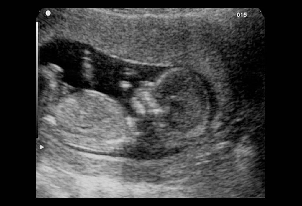
12 Week Pregnancy Ultrasound: Purpose, Process and Results
A 12 week pregnancy sonography is an essential first-trimester scan that confirms your baby’s growth, estimates the due date, checks for multiple pregnancies, and screens for certain chromosomal abnormalities. The World Health Organization (WHO) recommends at least one routine ultrasound before 24 weeks of pregnancy because it improves gestational age estimation, early detection of fetal anomalies, and overall pregnancy care (1). This makes the twelve week ultrasound one of the most important prenatal milestones.
In this article, you’ll learn the purpose of the scan, what happens during the procedure, how accurate the results are, and how to prepare for your appointment.
Why Do You Need an Ultrasound Scan at 12 Weeks?
Completing 12 weeks of pregnancy is an important milestone as it marks the completion of your first trimester, and your doctor may advise you to have an ultrasound scan to assess how your pregnancy is progressing. Here are a few things that your doctor will check in the 12 weeks ultrasound scan (2).
Like the previous scans, this scan helps to know the number of babies you are carrying, whether a single baby, twins or more.
It helps in establishing your baby’s gestational age by measuring the baby from crown to rump.
To check the foetal heartbeat.
To have a look at the baby’s arms, legs, chest, and head and assess if they are growing at a normal pace. true
The doctor will check the foetal spine as well to check for any abnormalities.
To measure nuchal translucency, which is the fluid under the foetal skin at the back of the neck.
The assessment of nuchal translucency helps in establishing any risk of chromosomal abnormality.
It will help the doctor to establish the health and the position of the placenta.
How to Prepare for Your 12th Week Scan?
You may be jittery and wonder what will happen during the scan. The first thing that you can do is to prepare yourself mentally for the scan. The cold and slimy gel may make you feel uncomfortable, but try to be calm and relaxed.
How Long Does This Ultrasound Scan Take?
You will be made to lie down in a peaceful room with dim lights. The entire scanning process at 12 weeks of pregnancy may take 15 to 30 minutes approximately.
How Is the 12th Week Ultrasound Scan Performed?
The doctor will apply a gel on your abdominal region and move the transducer device on your tummy. A black and white image will appear on the screen, and you can ask your doctor to explain it to you. It is normal to feel some pressure on your abdominal region, but it is not painful.
If you are overweight or your womb is too deep in your pelvis, then your doctor may recommend a vaginal scan.
What Can Be Seen in the Scan?
The following things can be observed about your baby by performing 12 weeks ultrasound scan (3):
The position of the mother’s placenta.
Baby’s limbs and spine.
Presence or absence of the foetal nasal bone and its length.
Urinary bladder and stomach of the foetus.
The blood circulation around the mother’s uterus region.
Your doctor can also spot twins in ultrasound at 12 weeks, which means if you are carrying more than one baby, the ultrasound scan will help your doctor determine the same.
How Does the Baby Look in 12-Week Scan?
At the 12-week scan, the baby is about 2 inches long, resembling a tiny human. Limbs, fingers, and toes are forming, with facial features becoming more defined (4). The scan provides an early glimpse of your growing miracle.
Can Down Syndrome Be Accurately Detected in the 12th Week Ultrasound Scan?
Wondering if you can detect down syndrome in 12 weeks pregnant ultrasound? Yes, if your baby is suffering from Down Syndrome, it can be detected in a 12-week ultrasound (3). If the baby has down syndrome, then there will be more accumulation of fluid at the nuchal fold, which is at the base of the baby’s neck. If your doctor suspects it, then the nuchal transparency test may be performed (5). Babies with Down Syndrome will have a thicker measurement in comparison to a healthy foetus.
What If Any Abnormalities Are Detected on the Scan?
The 12 week ultrasound 3D scan may be your first scan to establish the foetal growth and development. However, this scan also helps your doctor to assess any foetal abnormality. In case your baby is detected with any defect or abnormality, then you may be referred to a foetal medicine consultant. The consultant assesses the diagnosis made by your doctor and may ask you to undergo further tests or scans to confirm the abnormality.
Is This Scan a Part of Pregnancy Care?
Ultrasound scans are a great way of assessing your baby’s growth and development. These scans are affordable, non-invasive, and low risk. They are essential as they give you and your doctor a lot of information about your baby and you can also get 12-week ultrasound pictures. However, it is entirely up to the parents to take a call whether they want to go in for 12-week ultrasound scan or not. Most hospitals will give you the pictures of your ultrasound scan, and some of them may even give you a video of the scan. You can check with your doctor if this service is available.
Some parents are of the opinion that ultrasound scans should not be done. The main reason for this is that they will be put in a difficult position of making a decision to continue or abort the pregnancy, in case any foetal defect or abnormality is established.
Does Your Due Date Change at 12 Week Pregnancy Scan?
Indeed, your due date might be altered after the 12 week pregnant ultrasound. Statistics show that merely about 5% of infants are born exactly on their due date, particularly firstborns, who often arrive after the initially projected date.
Things to Keep in Mind
When you’re going for an ultrasound scan, remember to wear loose clothing so that it is easy for the doctor to access your belly.
If you have any questions for your doctor, writing them down would be helpful.
If you want a picture of the scan, you can contact the hospital in advance to ask if this service is provided.
Ask your doctor what kind of help is available in terms of prenatal classes or in case of an emergency. This can help you prepare for your baby’s birth and put your mind at ease.
FAQs
1. Where is my baby located at 12 weeks?
At 12 weeks of pregnancy, your baby is typically situated within the upper part of your uterus, nestled below your pelvic bone, and gradually moving upward as your pregnancy progresses.
2. Can my baby feel me at 12 weeks?
Yes, at 12 weeks, your baby may start feeling you soon.
3. Can a 12-week ultrasound confirm twins or multiple pregnancies?
Yes. A 12-week ultrasound can accurately confirm whether you are carrying twins or higher-order multiples. It also helps determine whether the babies share a placenta or amniotic sac, which is important for pregnancy management (6).
Previous Week: 11 Weeks Pregnant Ultrasound
Next Week: 13 Weeks Pregnant Ultrasound
Read more

18 Week Pregnant Ultrasound Scan
As you enter your 18th week of pregnancy, your doctor may recommend a detailed ultrasound examination to assess the growth and development of your unborn baby. An eighteen week ultrasound helps doctors monitor your baby’s growth, movements, and overall health. While for some women this may be the first look at their unborn baby, others may already have had an earlier scan.
An 18 week pregnant ultrasound can also provide clearer images of your baby, and some parents may even opt for an 18 week 3D ultrasound for a more detailed view. Here in the following article, we shall discuss all that you need to know about 18-week pregnancy ultrasound.
Why Should You Have an Ultrasound Scan at 18th Week of Pregnancy?
Ultrasound scan in your 18-20 week of pregnancy is a part of your routine check-up. Your doctor may advise you for an ultrasound scan because of the following reasons (1):
To establish the foetal age.
To check the number of foetuses (one or more than one)
To make estimates about the date of delivery.
To check the quantity of fluid around the foetus.
To check the cervix and the birth canal.
To see the position of the placenta
What Preparations Are Needed to Done This Scan?
There is not much preparation required for the 18 week scan. Drink sufficient water to keep your bladder full. This keeps your uterus in the correct place for the scan and helps your doctor to see the foetus clearly. Refrain from drinking too much water as it can make you extremely uncomfortable during the scanning process (2). You may ask your radiologist in advance as to how much water you are required to drink.
It will be a good idea to wear comfortable and loose clothes for the process so that the radiologist has easy access to your abdominal region.
How Long Does Eighteen Week Ultrasound Take to Complete?
On average, an ultrasound scan around 18-20 weeks of pregnancy may take from 30 to 45 minutes.
How Is This Scan Performed?
The radiologist will apply a gel on your abdominal region and run over a transducer over your belly. The scan is carried out in real-time, which means that what you will see on the screen, will be happening inside your womb during that time. You may ask the doctor to explain the images that are flashed on the screen. The main purpose of an ultrasound scan is to assess the foetal health and well-being (3).
What Can You See on 18 Week Scan?
You may ask your radiologist in advance if you wish you view the images of the scan on the screen. Though most hospitals or health centres provide for this facility, it is always good to inform in advance (3) (4) (5).
If you have an ultrasound scan for the first time, your doctor will tell you how many babies you are carrying and also your expected date of delivery.
You will also hear the foetal heartbeat for the first time.
Your radiologist may also show you your baby’s face, arms and legs. However, you may find it difficult to spot your baby’s organs, but that is absolutely fine because it is a bit difficult to understand the scan. You may ask the radiologist to help you understand the scan better.
The bones of the baby at 18 weeks pregnant ultrasound will appear white on the screen, the amniotic fluid will appear black, and the soft tissues will appear grey in colour.
After showing you the glimpses of the scan, the radiologist will then turn the screen towards himself and make other assessments. You will be handed a soft copy or a DVD of the scan after the process is over.
Are There Any Risks or After-effects of Ultrasound at 18 Weeks?
There are no after-effects of an 18 week anatomy scan, and you can easily resume your day to day activities. At the same time, there are no scientific pieces of evidence available that prove any ill-effects of an ultrasound scan on the expectant mother and her unborn child. There are no harmful radiations used to obtain the images, instead, very high-frequency sound waves are used for the purpose (6). In some cases where the foetus is farther away from the transducer, the process becomes little difficult and may take a bit longer. You may even be called after a few days for a re-scan. In the case of twins and multiple pregnancies also your doctor may face difficulties in establishing the foetal structures and you may be asked to come again after a few days.
Which Abnormalities Can Be Seen on the 18th Week Ultrasound Scan?
Some of these abnormalities may include the following (7):
Short limbs or absence of limbs
Cleft lip
Abdominal wall defects (protruding intestines)
Abdominal wall defects (protruding liver)
Abnormality related to the absence of top of the head
Severe kidney problem
Problems in the spinal cord
Excessive brain fluids
Chromosomal abnormalities
Severe heart defects
Any kinds of holes in the muscles separating the abdomen and the chest.
These medical conditions are not very common and rarely occur in babies. However, it is a good idea to get an ultrasound scan and rule out the possibilities of all these medical conditions.
What If There Are Signs of Problems on the Scan?
In case the radiologist is not able to see the foetus clearly in the scan, you will be called at a later date for a re-scan. In case you are obese, or the baby is tucked too deep into the womb, a re-scan may be suggested too. However, if your doctor sees any kind of above-mentioned abnormality or other defects, you will be told about them. You will also be told to get an appointment with a foetal medicine expert to know about the intricacy of the defect and the options available to you (5).
In case of serious abnormalities, you may be suggested to terminate the pregnancy. However, some abnormalities may require immediate medical attention soon after birth.
What to Expect After 18 Week Ultrasound?
An 18 week fetus ultrasound does not always provide a confirmed diagnosis for medical conditions, but it helps doctors identify whether any additional evaluation may be needed. After reviewing the scan, your healthcare provider will discuss the results and check if your baby’s growth and development are progressing normally.
If the doctor notices any unusual findings or potential concerns, they may recommend further testing or a follow-up ultrasound with a maternal-fetal medicine specialist (2). In some cases, additional diagnostic procedures such as amniocentesis may also be advised to gather more detailed information about the baby’s health (5).
FAQs
1. What can be seen in an 18 week 3D ultrasound?
If you are 18 weeks pregnant, 3D ultrasound may provide more detailed and lifelike images of your baby’s facial features, hands, feet, and body structure compared to a standard ultrasound.
2. Can twins be detected during an 18 week ultrasound?
Yes, an ultrasound at 18 weeks can clearly confirm a twin or multiple pregnancy and help monitor the growth and development of each baby.
3. What if my baby is not moving during the ultrasound?
Sometimes babies may be sleeping during the scan. The technician may ask you to change positions or move slightly to encourage movement.
An ultrasound scan at 18 weeks is a great way of knowing about your unborn baby’s health and well-being. Where some parents are too eager for the scan, others may decide not to go for the scan. It is absolutely left up to the parent’s discretion, whether or not they want an ultrasound scan. You may talk to your doctor about your options.
Previous Week: 17 Weeks Pregnant Ultrasound
Next Week: 19 Weeks Pregnant Ultrasound
Read more
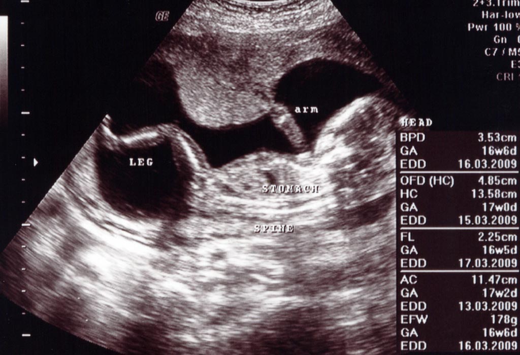
16 Weeks Pregnant Ultrasound : What to Expect?
Hey, it’s your sweet 16th month of pregnancy! You are beginning your fourth month and entering the second trimester. There’s a lot happening inside your body, and your doctors mean well for you if they suspect anything serious. About 1 in 100 babies is born with a heart defect. Don’t worry; early interventions, like a 16-week ultrasound, can spot issues early. While 16-week ultrasounds aren’t routine, the possibility of a heart defect isn’t just one reason your doctor would recommend you have the 16-week scan. If you are scheduled for one, you will likely have it transabdominally to help the doctor assess the baby’s development, including growth of the torso, limbs, head, and facial features. In this article, we discuss everything crucial you need to expect from your ultrasound at 16 weeks. Read on!
Why Should Pregnant Women Have an Ultrasound at 16 Weeks?
Things start to get a bit exciting once you hit the 16-week mark, and an ultrasound is important to understand your baby’s progress. Prenatal care is an integral part of every mother’s journey, and here are a few reasons as to why an ultrasound is necessary at this stage.
It can help you understand your baby’s growth, and the sonographer can help measure certain parts of your baby’s body to assess any concerns.
Ultrasound will become an integral part of your journey into motherhood. It helps determine the size, structure and any pathological lesions on the foetus (1).
It can present a visual representation of your baby and help prepare for routine and emergency prenatal care.
It can help understand if your baby is healthy and safe and if there are any abnormalities.
Genetic issues can also be determined, such as Down Syndrome or any other chromosomal defects, or have any other neural tube defect.
The 16-week foetus ultrasound can determine the heartbeat and health of the heart and other organs. It can help determine if there is any heart defect as well.
What Preparation Is Needed for a 16-week Scan?
Your baby at 16 weeks’ ultrasound is the largest it has been in your pregnancy to date, and a few things need to be done before you go for a 16-week scan. Your baby, which measures around four and a half inches during the 16th week, will grow double in size very quickly, month by month, from here onwards. There will definitely be a weight gain during this time, but morning sickness will completely disappear.
Your body will start producing milk, which will lead to breast augmentation. In addition, the belly will be larger, and the skin colour will change to some extent as well.
Before a scan, it is important to carry documents about your and your partner’s medical history to your gynaecologist and ask questions regarding any symptoms or issues you may be experiencing. You should also get a full physical examination, including thyroid, breasts, lungs and heart. You should get a lab test done to check signs of gestational diabetes, preeclampsia or any other urinary tract infections through urine tests.
Familiarising oneself with terms which are associated with pregnancy and medical procedures, such as ultrasound, will also help in understanding your baby better.
How Long Does a Sixteen-Week Ultrasound Take to Complete?
An ultrasound can last anywhere between 30 and 60 minutes. Before an ultrasound, a patient is asked to drink lots of water or fluids and stay away from solid food. You cannot access the toilet when you wait for the scan, but you can go relieve yourself once the procedure is complete (2).
Before you begin the test, make sure to prepare yourself mentally to wait and to understand that they take time, to image the sonography. Imaging reports also take a day to be produced, but both parents will be shown the image of their baby on the screen.
How Is Sixteen Week Scan Done?
A 16-week foetus ultrasound is done by using a device that emits high-frequency sound waves into the body. The waves hit and bounce off the various organs of the body as they travel. A probe picks up these echoes created by the bouncing sound waves and starts converting them into a moving image. These images can be accessed through a graphics display monitor. These tests are usually conducted in clinics, diagnostic centres or the radiology section of a hospital. A specially trained expert will be the only one who will be able to conduct a scan.
What Can Be Seen in the Scan?
When you get an ultrasound done at 16 weeks, your belly will look like a small cantaloupe. On the inside, however, your baby is around 4-5 inches in size and will continue to grow. Hair and fingernails are starting to develop, although they will be more noticeable once you progress in your pregnancy. You will be able to make out the head, the eyes, ears and torso of your baby. All the muscles and bones are in place, and the central nervous system is starting to exert control, so you may even see your baby move during an ultrasound.
Your baby is beginning to make expressions, but any smiling or frowning will happen randomly, as they lack muscle control at this stage. The nervous system is still maturing, allowing your baby to start moving their arms and legs. Your baby’s hands can clench into fists, and they might start jabbing around inside you as well (3).
Is It Possible to Determine the Gender of the Baby at a 16 Week Ultrasound?
Yes, you can ascertain your baby’s gender during the 16-week gender scan. The determination should be reliable since the baby’s external anatomy is fully developed by then.
However, it’s important to note that accuracy may be compromised by the baby’s positioning, potentially hindering the physician or ultrasound technician from obtaining a clear view of the baby’s anatomy for gender confirmation.
Disclaimer: Under the Pre-conception and Pre-natal Diagnostic Techniques (Prohibition of Sex Selection) Act, 1994, (PC & PNDT Act), determining or disclosing the sex of a fetus before birth is illegal in India, except for specific medical indications permitted by judicial law. Therefore, all healthcare providers, including doctors, clinics, and hospitals, in India cannot reveal a baby’s gender during a 16-week fetus ultrasound or at any stage of pregnancy (4).
What If Any Abnormalities Are Seen in the Ultrasound?
When you go for an ultrasound, the sonographer will also conduct a variety of other tests to determine any abnormalities in your baby’s growth such as measuring the length of the baby’s nasal bone to determine any abnormalities in growth is one. There are several blood tests which are offered to determine any risks or abnormalities that can arise at this stage of the pregnancy.
The Triple screen is a prenatal blood test that will help assess any issues with the baby. The test primarily helps test levels of alpha-fetoprotein (AFP), a protein produced by the foetus’s liver and two hormones: human chorionic gonadotropin (hCG) and estriol, to determine any birth defects such as Down Syndrome, Spina Bifida (5).
It is important to discuss any concerns or anything that comes up during an ultrasound with your doctor in order to determine the next course of action for you and your baby. It is also important to take any medication or tests that are required to keep you and your baby healthy.
FAQs
1. Is a 16-week ultrasound safe for my baby?
Yes. Diagnostic ultrasounds use sound waves rather than radiation and are considered safe when performed by trained healthcare professionals for medical purposes. Thus, an ultrasound in a 16-week pregnancy is safe.
2. Can I expect twins at a 16-week ultrasound?
Yes, a normal 16-week ultrasound can show babies in detail if you are carrying twins. You can later have a 16-week twin ultrasound to confirm.
3. Which test can be done at 16 weeks?
Between 16 and 18 weeks of pregnancy, your doctor may provide you with a second-trimester screening test (16 week 3d ultrasound or normal ultrasound), known as the multiple marker test or triple screen.
4. Can a 16-week ultrasound detect birth defects?
Some structural abnormalities may be visible at 16 weeks, but the detailed anatomy scan performed around 18–22 weeks is usually more effective for identifying congenital conditions.
At 16 weeks every mother will have the pregnancy glow and move into the second trimester which is an enjoyable phase. As your baby continues to grow, more movement from the foetus will show, the belly will grow bigger, and the body prepares for the next phase of motherhood.
Previous Week: 15 Weeks Pregnant Ultrasound
Next Week: 17 Weeks Pregnant Ultrasound
Read more

11 Weeks Pregnant Ultrasound: Picture, Procedure, & more
An 11th-week ultrasound is a common thing during a pregnancy check-up to determine your due date. By around eleven weeks, your baby officially becomes a foetus from an embryo. Distinct facial features and genitalia are developing in your baby, and the eleven-week scan gives an exciting preview of the way your baby is growing inside you. The 11 weeks pregnant ultrasound is also medically significant for ruling out any anomalies, syndromes, or malformations. Let’s move ahead and see what the scan performed at eleven weeks includes, what an 11-week-old fetus looks like, why you might need an 11 weeks fetus ultrasound, and how it is done.
Purpose of the 11 Week Ultrasound Scan
The first-trimester pregnancy scan is always very special as it gives a glimpse of the developing baby. The week 11 scan gives you a sneak peek at how your baby looks for the first time. It is also the earliest stage at which the doctor can pick up certain anomalies in the baby.
At this stage, you likely won’t notice much detail regarding your baby’s limb and organ development. Because of this, it’s quite possible that you won’t have an ultrasound at 11 weeks pregnant. However, there are some reasons why you might have one.
The purpose of the week 11 scan is manifold. Here are some reasons why the 11-week scan should not be missed.
During the 11-week scan, the foetus is screened for chromosomal abnormalities like Down’s syndrome. The scan results are evaluated along with the blood test results to arrive at a diagnosis.
The position and size of the placenta are checked.
The amount of amniotic fluid is assessed.
The nuchal translucency (NT), the translucent space beneath the foetus’s neck, is used as a risk factor for the syndrome. Nuchal translucency is also used as a guideline to rule out any congenital cardiac disease.
Multiple pregnancies, if any, are clearly visible during this scan. Twins are usually visible at this stage as two clearly separate gestational sacs.
A general pregnancy health assessment is made.
The gestational age of the baby is checked based on the growth of the fetus.
Check for an ectopic pregnancy.
How to Prepare for Your 11 Week Scan
As the uterus lies low in the abdomen, going with a full bladder can help provide better visualisation of the foetus. You can drink a few extra glasses of water and prepare yourself for the scan. However, some sonographers prefer a partially full bladder. Wearing comfortable, loose clothing that easily exposes the abdomen can facilitate the procedure.
How Long Does the 11 Week Ultrasound Take?
The first-trimester ultrasound at eleven weeks is usually a simple procedure that takes a few minutes. An experienced sonographer measures the nuchal translucency quite easily. They also check for other vital growth parameters, such as crown-rump length, the diameter of the gestational sac, placental position and size, etc. The position of the foetus can delay the scan. Sometimes the baby may be turned to one side or curled up, making it very difficult to measure the tissue below the neck. Usually, the sonographer suggests repeating the scan at a later time until the correct foetal position is achieved.
How Is the 11 Week Ultrasound Scan Performed?
The week 11 scan is usually performed trans-abdominally. A cooling gel is applied to the lower abdomen, and the transducer device that sends ultrasound waves into your uterus is moved over the tummy to get a clear view of the foetus. The baby’s images are taken from the top and sides to provide a better view of all internal and external organs.
What Can Be Seen in the Scan?
Many mothers are anxious about the 11-week ultrasound. Although it is a very crucial scan that rules out any abnormalities in the foetus, there is no need to panic. It is probably the first time you get to see your moving baby, so be prepared for a lot of excitement.
The foetus is about the size of a fig (about 1.57 inches long).
The baby, at 11 weeks, looks more human with the well-formed nose, lips, webbed fingers and toes, and thin limbs (1).
By the end of the 8th week, bones begin to form, and by the 11th week, the baby’s head and bones have developed and are getting stronger (2). They will be pronounced in the 12th week.
The heart is well-formed and pumping well, and the lung tissues are in development with trachea and bronchi, and some arteries and capillaries are already developed (3).
The body is still disproportionate, with a large head and a comparatively small torso.
Facial features are more prominent now (4).
Their skin is translucent, and the eyelids are closed (5).
You can catch your baby bouncing and kicking in the watery amniotic sac.
The baby’s genital organs are beginning to form this week and may not be clearly visible in the scan.
The foetus at this stage is only an inch or two long but has a rapidly developing organ system.
What Should a Fetal Heartbeat Be At an 11 Week Ultrasound Scan?
At 11 weeks, your baby’s heartbeat is expected to rise above 170 bpm, increasing from 110 bpm around 5 to 6 weeks (6). An 11 weeks pregnant scan may provide this information, although some early ultrasounds might detect a heartbeat as soon as six weeks.
However, you can typically expect to hear a heartbeat using a Doppler fetal monitor around 10 weeks.
Although your developing baby might be evaluated for an irregular heartbeat (fetal arrhythmia) between 12 and 14 weeks, it’s more likely to be detected during the 20-week assessment (7).
What If Any Abnormalities Are Seen in the Scan?
If the 11-week foetus ultrasound indicates the possibility of chromosomal abnormalities, then you can talk to your doctor about the other confirmatory tests you can take. The NT values reflected in the scan are not conclusive evidence for a foetal abnormality. A blood test is often performed at the same time as an NT scan to detect trisomy 21 (Down’s syndrome) and trisomy 18, and sometimes 13, to corroborate the scan’s findings (8).
The doctor may also suggest an invasive procedure called amniocentesis to check for the DNA of the foetus. Chorionic villus sampling (CVS) can also be offered earlier (11–13 weeks). There is a slight risk of miscarriage, and couples can decide to take it after discussing it with their genetic counsellor or doctor (9). To avoid the invasive procedure, you can wait for the anomaly scan at weeks 18-20, which can detect some mild indicators of abnormal growth. Based on all these indicators, the doctor or genetic counsellor will help you with the diagnosis and advise you about the course of action.
The week 11 NT scan is crucial in terms of identifying any congenital anomalies in the foetus. Although the scan result alone is not enough for a diagnosis, it definitely serves as a guideline for further tests.
Previous Week: 10 Weeks Pregnant Ultrasound
Next Week: 12 Weeks Pregnant Ultrasound
Read more

Impact of Birth Method on Long-Term Immunity: C-Section vs. Vaginal Delivery
As parents, we want to do everything possible to give our babies the best start in life. We think about feeding, sleep, and care, but one thing we don’t always realise is that the way a baby is born can also affect their immune system. Surprising, right? When a baby is born, the focus is often on the first cry, tiny fingers, and the joy in the room.
But little do most people know the method of delivery can have a lasting impact on the baby’s health. The way your baby enters the world plays a bigger role in their immunity than many might think. Keep reading to find out how the birth method affects your baby’s immune system and how you can support them in the best way possible.
Vaginal vs. C-Section Delivery
During a vaginal birth, babies pass through the birth canal and pick up beneficial bacteria from their mother (1). These friendly microbes start building the baby’s gut microbiome; a community of bacteria that helps with digestion, nutrient absorption, and, most importantly, training the immune system (2).
Breastfeeding adds another layer of support. Breastmilk contains prebiotics, which are special fibres that feed the good bacteria in the baby’s gut, helping them grow strong and multiply (3).
Together, this early exposure and nutrition helps to shape a baby’s natural defences from day one.
However, babies born via C-section enter the world without passing through the birth canal. That means they do not come in contact with the first natural bacteria’s present in the mother vagina (4). Instead, their early gut bacteria may be similar to the bacteria present in the the hospital, which can sometimes mean less good quality bacteria in the gut.
This difference doesn’t mean anything is “wrong”; but it may influence how the immune system matures.
Why the Gut Microbiome Matters for Immunity?
Inside all of us is something called the gut microbiome, a collection of bacteria that lives in our intestines. These bacteria help us digest food, protect us from infections, and strengthen our immune system (2) (5).
For babies, the gut microbiome starts forming even before birth. A strong and healthy microbiome helps build a stronger immune system and may even reduce the risk of allergies, asthma, and other illnesses later in life.
How C-Sections May Affect Immunity?
Babies born by C-section may have a slower start in building their gut bacteria, and that might affect how their immune system develops. Some studies suggest that babies born via C-section may have a slightly higher risk of developing allergies, asthma, and possibly infections early in life (6).
But this isn’t something to stress about. We can support their gut and immune health through smart nutrition, especially with Synbiotics.
How Do We Support the Infants Born via C-Section?
In such cases, breastmilk plays a key role. It provides not just key nutrients like fats, proteins, carbohydrates, vitamins and minerals, but also good bacteria (probiotics) and food for those good bacteria (prebiotics) which help to build gut health and also boost immunity. This is especially important for babies born via C-section. Breastmilk, is naturally rich in Synbiotics and together, they create a healthy gut environment for the baby, making digestion easier and helping the body fight infections better. Research shows that including Synbiotics in a baby’s diet at the right time can support their overall health, by boosting their immune system.
What Are Synbiotics?
Synbiotics are a powerful combination of probiotics (the good bacteria) and prebiotics (the food that helps them thrive). When taken together, they work in harmony to support a healthy gut and a strong immune system (7). For babies born via C-section, Synbiotics can help bring their gut microbiome closer to that of babies born through vaginal delivery (8).
Specific Synbiotic combinations help mimic the benefits of naturally available bacteria. These Synbiotics can be especially helpful for babies born via C-section or those with sensitive digestive systems.
While Synbiotics aren’t a replacement for the natural process of birth, they can be a valuable way to help bridge the gap, giving your baby’s immune system the support it needs during those early, important months. If your baby was born through C-section, you might want to ask your doctor about how and when to introduce Synbiotics for better gut health and build strong immunity.
Well, not every parent can choose how their baby is born, and that’s completely okay. What matters is how we support them after birth. With the right nutrition, care, and a little help from science, your baby’s immune system can grow stronger every day.
References/Resources
1. American Museum of Natural History – Building Your Microbiome from Birth
2. Cleveland Clinic – Gut Microbiome
3. PubMed Central – The Prebiotic and Probiotic Properties of Human Milk: Implications for Infant Immune Development and Pediatric Asthma
4. National Library of Medicine – Vaginal Delivery
5. Harvard T. H. Chan School of Public Health – The Microbiome
6. PubMed Central – The infant microbiome development: mom matters
7. PubMed Central – Synbiotics: a technological approach in food applications
8. PubMed Central – Clinical outcomes following pre-, pro- and synbiotic supplementation after caesarean birth or antibiotic exposure in the first week of life in term born infants: A systematic review of the literature
Read more
Sort By
 Latest
 Most Popular
 Trending
Please select atleast one Filter