

















.svg)
.svg)
.png)


Complications
Suggested articles for you

Uterine Rupture – Causes, Symptoms and Treatment
If you’ve heard someone you know had a uterine rupture, we are sure it took some time for the news to sink in and also raised concerns and curiosity about what it really is. The name itself sounds like a nightmare, and before you read further, you must know that yes, it is scary, but it is a very rare condition. Though you may have one or more risk factors that are mentioned below, the chance that you might face this is low. Studies show that a majority of women who deliver after a prior caesarean have a safe and normal birth. According to the National Health Institute, 992-993 women out of 1,000 give birth without the complication of a uterine rupture.
Let’s dive deep into the topic and know what uterine rupture really is.
What Is Uterine Rupture?
Uterine rupture or a tear in the uterine wall during pregnancy is a very rare complication usually observed in the last trimester of pregnancy which can cause maternal and foetal distress.
The uterus is a stretchable bag-like structure located in the abdominal cavity, and its wall is three-layers thick. These layers are –
The inner, thin layer called the endometrium, which is the most active layer responding to all the hormonal changes and is highly specialised.
The middle layer, a thick layer of muscles, which forms the main bulk and gives the strength to the wall of the uterus.
The outermost layer called the serosa or the peritoneum, which is a very thin layer that forms a coat or an envelope and hence separates the uterus from the abdominal cavity.
Uterine rupture occurs when the uterine wall is disrupted in such a manner that the contents of the uterus (like the foetus, the placenta, and the umbilical cord) can be expelled into the abdominal cavity.
On the other hand, uterine scar dehiscence or uterine window or scar separation involves the disruption and separation of a pre-existing uterine scar, and it does not disrupt the serosa. Hence, the contents of the uterine cavity remain within the uterus.
Uterine scar dehiscence is far more common than uterine rupture and only rarely results in major maternal or foetal complications. Although a uterine scar is a well-known risk factor for uterine rupture (most of which arise from prior cesarean delivery), the majority of the events involving the disruption of uterine scars results in uterine scar dehiscence rather than uterine rupture. These two conditions need to be clearly distinguished, as the clinical management and the resulting clinical outcomes differ significantly.
Uterine Rupture & Pregnancy
Uterine rupture usually occurs during peripartum, that is the time period shortly before, during, and immediately after delivery. This is obviously because it is the time when the foetus has attained the maximum size, and the uterus is maximally stretched. It can either occur in women with a native, unscarred uterus or a uterus with a surgical scar from previous surgery done for any reason – previous Cesarean section or any other surgery like a myomectomy (partial removal of the diseased uterine wall be it by open surgery or a laparoscopy).
What Are the Chances of the Uterus Getting Ruptured?
Even in high-risk groups, the overall incidence of a ruptured uterus during delivery is quite low. It occurs at the rate of 1 in 1,146 pregnancies (0.07%). The chances of uterine rupture are even fewer in an unscarred uterus. But if you have undergone a previous Cesarean section or any surgery on the uterus, the risk of a uterine rupture during delivery doubles consequently. Also, the higher the number of various risk factors present, the more is the chance of rupture, the incidence still being very low.
What Are the Risks for a Uterine Scars Separation?
The possible causes of the uterus rupturing are different for an unscarred uterus and a scarred uterus, the latter being accountable for most of the cases.
Reasons for Uterine Rupture in an Unscarred Uterus
An unscarred uterus is least susceptible to a uterine rupture. The incidence is only 0.012% that is 1 in 8434 pregnancies – almost all the cases being those of second or third pregnancies rather than first pregnancies. So, if this is your first delivery and if you have never had any surgery done on your uterus, you need not worry.
Following are the risk factors in an unscarred uterus:
Grand multiparty (several pregnancies): Studies say that the chance of rupture is more if there have been more than three deliveries in the past. In successive deliveries, there is a progressive thinning of the uterine wall and hence the risk increases.
Obstructed labour: This occurs when a woman is allowed to go in labour (known as the trial of labour) in order to have a natural vaginal delivery, but the labour is aborted, and a C-section is performed to complete the delivery. Doctors typically take this decision is the birth canal is too small for the baby to be born.
Neglected labour: This usually happens when there is no access to medical care, or in the case of home-births.
Malpresentation: In this case, a different body part of the baby appears first during the birth instead of the head. Usually, the head is the ‘presenting’ part of the baby being born; however, sometimes the child’s presenting part is the hip or the shoulder or the face or the legs. This causes abnormal and uneven pressure over the uterus, which then gives away.
Uterine instrumentation by the doctor during difficult labour (such as the use of forceps) may cause uterine ruptures.
Labour induction or augmentation using oxytocin (a drug used to increase uterine contractions) can increase chances of uterine rupture.
Congenital abnormality of the uterus.
Reasons for Uterine Rupture in a Scarred Uterus
A single cesarean scar increases the overall rupture rate to 0.5%, with the rate for women with two or more cesarean scars increasing to 2%. Medical literature suggests that there is an increase in the complications following vaginal birth after previous cesarean section (VBAC) due to uterine rupture. This and the medico-legal fears is the reason why there is a decline in the doctors offering and women accepting planned VBAC in the UK and North America and now also in urban India. Can uterine fibroids rupture? Well, the scar of myomectomy on the uterus (the surgery done to remove the fibroids) can give way to uterine rupture. The fibroids per se, causing a rupture in an unscarred uterus is extremely rare.
Risk factors in a scarred uterus:
1. Trial of Labour after Previous C-Section (TOLC)
There is a threefold increased risk of spontaneous uterine rupture after a previous C-section compared with women who undergo elective repeat cesarean delivery. But, it is quite interesting to note that if there has been one successful VBAC (vaginal birth after C-section) earlier, the chance of uterine rupture during VBAC subsequently is decreased.
Multiple potential theories exist, but the two most obvious are that a successful prior VBAC attempt ensures that –
The maternal pelvis is tested and is adequate to permit passage of the fetus.
The integrity of the uterine scar has been tested previously under the stress/strain conditions during labour and delivery that were adequate to result in vaginal delivery without prior uterine rupture.
2. Type of C-Section Scar
The upper vertical scar in olden times is now almost obsolete as it carried the maximum risk of rupture. The lower transverse scar most commonly used today is least susceptible to rupture.
3. Interdelivery Time Interval
There is a threefold increased risk of a rupture if the interdelivery time between the previous C-section and the subsequent delivery is less.
4. Maternal Age
There is a threefold increase in the rate of rupture if the maternal age is greater than 30 years compared to younger mothers.
5. Twins
There is no increased risk in twin pregnancy compared to singleton pregnancy.
6. Size of the Baby
There is a slightly higher risk of rupture if the weight of the baby is more (> 4000 gms).
7. Advanced Gestational Age
There is a significantly higher risk of rupture if the gestational age is greater than 40 weeks as usually they are associated with induction of labour.
Signs & Symptoms of Uterine Rupture
What happens if a scar starts to tear during labour? The initial signs and symptoms are quite nonspecific, and that makes the diagnosis difficult, delaying the definitive treatment. The signs and symptoms may vary from being minimal like in small dehiscence to a severe life-threatening one, like in the case of a large rupture which involves a major blood vessel. It largely depends on the timing, site, and extent of the uterine defect. Uterine dehiscence at the site of a previous uterine scar is typically serious because the scar has relatively fewer blood vessels.
What the Mother Can Experience
A mother, when in labour, or due to lack of experience, may not be able to notice any symptom and hence delay the diagnosis and treatment. Following are some symptoms that may indicate a uterine rupture:
Abdominal pain: Sudden or a sharp pain, which the mother can differentiate from normal labour pain only if very alert and informed. This pain persists even between the contractions. A woman undergoing a painless delivery under spinal anaesthesia will not feel the pain. Thus, abdominal pain is an unreliable and uncommon symptom of uterine rupture.
Can no more feel the uterine contractions or lesser intensity contractions.
Sharp onset of pain at the site of the previous scar.
Excessive vaginal bleeding.
Giddiness and loss of consciousness.
What a Doctor Can Pick Up
Foetal distress: The most common sign and often the first and only sign of uterine rupture is an abnormal foetal heart rate as measured by electronic foetal monitor. This sign is usually present in 79-87% of patients. Hence, it is very important for the doctor to monitor the foetal heart rate when a mother is undergoing labour.
Diminished baseline uterine pressure.
The recession of the presenting foetal part – the baby may sometimes slide back into the uterus as the uterus can no longer push the baby outwards (on account of getting ruptured).
Excessive vaginal bleeding.
Shock: This will occur after there is a lot of blood loss and a drop in the blood pressure.
These signs are not specific for uterine rupture, but only indicative and needs a proper diagnosis by experts.
What Happens If the Scar Starts to Tear During Labour?
The consequences of uterine rupture depend on its severity, the time between diagnosis and treatment and the level of medical care available.
Foetal consequences are admission to neonatal intensive care unit, fetal hypoxia or anoxia (decreased or no oxygen to the baby respectively), and neonatal death. The chance of death varies from 6-17%, but the incidence of perinatal death associated with uterine rupture is decreasing in the modern era.
Maternal consequences are bleeding, low blood pressure, bladder injury, need for hysterectomy, and maternal death.
Diagnosis of Uterine Dehiscence
Uterine rupture is an emergency, and it needs rapid diagnosis and treatment. Because of the short time available before the onset of irreversible damage, time-consuming diagnostic methods and sophisticated imaging modalities have only limited use. It is only the clinical expertise of the doctor that will help based on the standard signs and symptoms.
One may wonder – is it possible to predict uterine rupture well in advance and be prepared? It is, but only in selected few cases. Various diagnostic techniques have been used to attempt to assess the individual risk of uterine rupture in selected patients. MRI may aid in evaluating the status of uterine scar in an affected woman. Ultrasonography may be useful for detecting uterine-scar defects after caesarean delivery by measuring the thickness of the uterus wall. The risk of uterine rupture after previous caesarean delivery is directly related to the thickness of the lower uterine segment, as measured during transabdominal ultrasonography at 36-38 weeks of gestation. The risk of uterine rupture increases significantly when the uterine wall is thinner than 3.5 mm.
Uterine Rupture Treatment for Pregnant Women
The most critical part of the management of uterine rupture is timely diagnosis and commencement of the treatment as fast as possible. As a rule, the immediate stabilisation of the mother (including fluid resuscitation and blood transfusion) and the delivery of the foetus within 10-37 minutes of uterine rupture is necessary to prevent serious complications to both the mother and the child. The newborn may require admission to the NICU (Neonatal Intensive Care Unit) and observation. Later, the mother might need a surgical intervention which again may vary from a simple tear repair to removal of the uterus (hysterectomy) depending on the type and the extent of uterine rupture, the degree of bleeding, the general condition of the mother and the mother’s desire for future childbearing.
Can Uterine Rupture Harm the Baby and the Mother?
Yes. Uterine rupture can cause significant harm to the mother and the baby. It can also result in death. But in a well-managed case with prompt diagnosis and treatment in a well-equipped hospital, the mother and the baby usually do well, and death is an extremely rare occurrence. In some cases, uterine rupture recovery time is prolonged and needs longer hospital admission adding to the cost. Uterine dehiscence is less harmful and in fact, may not cause any trouble at all.
How Can Uterine Rupture Be Prevented?
The most direct prevention strategy for pregnancy-related uterine rupture is to minimise the risk factors. The risk modification begins even before the first conception until the last delivery.
What Can a Mother Do?
Plan pregnancy before the age of 30 years.
Don’t opt for a Caesarean section only because you want to avoid the painful labour. Natural is always the best. A c-section scar is not natural.
Spacing is important. Avoid pregnancy within two years of a Caesarean section.
Be a good patient. Timely consultation as advised by your doctor, especially in the last trimester and close to the expected date of delivery helps.
Feel free to consult when something unusual is felt like sudden abdominal pain, loss of contractions decreased foetal movements, etc.
Crossed your expected date of delivery and aren’t getting labour pains yet? Meet your doctor immediately.
What Can a Doctor Do?
Identify a patient at high risk of uterine rupture.
Try to predict the event with the help of ultrasonography or MRI.
Counsel and inform the mother in detail regarding her condition and help take the best decision.
Take timely decision about whether to conduct a trial of labour or opt for a Caesarean section depending on the risk factors.
This should be done – Tight closure of the c-section wound in at least two layers and avoiding a single layer stitch.
Close monitoring of the foetal heart rate and foetal activity is important.
Prompt diagnosis of the uterine rupture and treatment is crucial too.
Although uterine rupture can have dire consequences, it is important to remember that it is extremely rare, and can be harmless if detected early. Being aware and staying alert are the best ways of avoiding uterine rupture.
Read more





Calcified Placenta in Pregnancy: Causes, Diagnosis, and Prevention
Key Takeaways
Placental calcification is usually a natural part of placental aging, especially near the end of pregnancy.
Early or extensive calcification may require closer monitoring of your baby’s growth and well-being.
Ultrasound is commonly used to identify and assess placental calcification.
There is no proven way to prevent normal placental calcification, but healthy pregnancy habits can support overall maternal and fetal health.
A calcified placenta means calcium deposits have developed in the placenta, often as part of the natural changes that occur as pregnancy progresses. In many cases, these deposits simply reflect a normally aging placenta, especially toward the end of pregnancy. However, when calcification appears earlier than expected or is more extensive, your doctor may recommend closer monitoring of your baby’s well-being.
But when should placental calcification raise concern, and what causes it to happen earlier? This article covers the possible causes and risk factors, how calcification is diagnosed on ultrasound, what it may mean for your pregnancy, and whether it can be prevented.
What Is a Calcified Placenta?
A calcified placenta refers to calcium deposits that develop within the placenta as it matures during pregnancy. This is a natural change that typically becomes more noticeable as the due date approaches, reflecting the placenta’s normal aging process. In most cases, late-pregnancy calcification is a common finding and does not indicate a problem with the pregnancy (1).
When Does Placental Calcification Occur?
Placental calcification can start as early as the 12th week of pregnancy and may gradually increase as the placenta matures. The timing and extent of these calcium deposits are commonly described using four stages, from stage 0 to stage 3 (2).
Stage 0: Before 18 weeks of pregnancy
Stage 1: 18-29 weeks of pregnancy
Stage 2: 30-38 weeks of pregnancy
Stage 3: Around 39 weeks of pregnancy
What Causes Placental Calcification?
Calcification of the placenta is quite common, with over half of all placentas showing some level of calcification when a pregnancy reaches full term. Some of the possible causes for the calcification of placenta include (3):
Smoking
Hypertension
Bacterial presence in the placenta
Placental abruption
Unexpected reaction to supplements or medication
Exposure to radiation or low-frequency noise
What Are the Symptoms of a Calcified Placenta?
The most common symptoms indicating placental calcification include less or no movement of the baby, even though they are close to term. We recommend seeking medical advice immediately if you notice any of the following (4):
If you haven’t felt any movement from your baby by the time you reach 24 weeks of pregnancy.
If your baby’s movements are noticeably reduced compared to their usual activity.
If, during the third trimester, your baby moves less than ten times within a span of 1-2 hours.
How Is the Age of the Placenta Calculated?
Estimating the exact age or calcification of the placenta is difficult. Trained radiologists who conduct ultrasounds may be able to tell you the approximate age of the placenta through the ultrasound images. However, different doctors may have different interpretations of the same image.
How Is Placental Calcification Diagnosed?
The solitary way in which we can diagnose placental calcification is through pelvic ultrasonography (5). However, in many cases, it gets diagnosed during the routine sonography that is conducted during pregnancy.
How Does Placental Calcification Affect Delivery?
Placental calcification near the end of the pregnancy period is termed normal. In fact, the start of placental calcification could mean that you are nearing your delivery time. However, calcification before the 37th week or the maturity of the foetus can pose a problem for the baby. It can cause low birth weight, preterm birth and, in rare cases, the death of the foetus (6). However, with immediate medical intervention and continuous monitoring of the mother and baby, it can be taken care of. Let’s understand placental calcification at different weeks and what it implies.
1. Calcification of Placenta Before 32 weeks
If calcification starts before the 32-week gestation, it is termed as early preterm placental calcification.
The start of calcification at this period of pregnancy can be extremely dangerous for the mother as well as the baby (7).
The mother may experience postpartum haemorrhage and placental abruption.
The baby may be born preterm and may experience all types of risks associated with premature births.
The baby may have a very low Apgar score and a low birth weight.
In rare cases, the foetus may die in the mother’s womb itself.
2. Calcified Placenta Between 28 and 36 weeks
Pregnant women, who suffer from hypertension, diabetes, kidney disorders, anaemia, cardio problems etc., or in other words, women with high-risk pregnancies, are more at risk or danger than women with low-risk pregnancies or normal pregnancies.
It requires continuous monitoring of the foetus and the mother by doctors.
Calcification at this stage of pregnancy could result in adverse outcomes for the mother as well as the foetus.
3. Placental Ageing at 36 weeks
Grade III placental calcification can induce hypertension during pregnancy which can prove to be fatal for the foetus and result in pregnancy-related complications for the mother.
The baby born in this condition will probably have a low birth weight.
The mother has to have a preterm delivery which will in all probability, be through C-section.
4. Placental Calcification from 37 to 42 weeks
Delivery from the 37th week onwards can be termed as safe. The foetus is fully matured and may not face any risks.
The weight of the baby is usually normal.
Doctors do not term it as risky for the mother or the baby.
However, babies should be delivered before the 42nd week since placental calcification makes the placenta incapable of delivering nutrition and oxygen to the foetus. An inadequate supply of oxygen could also pose a risk to the baby’s brain and damage it.
Can Early Placental Calcification Affect the Baby?
Premature placental calcification may pose several complications for the mother and baby. Since it is a condition wherein deposits of calcium form in the placenta and obstruct the flow of oxygen and nutrients to the foetus, it can be termed as a dangerous condition. As the baby does not receive the required amount of nutrients or oxygen, it has to be delivered preterm, which results in low birth weight. Due to the calcification process, some parts of the placenta become dead and don’t function normally. The obstruction of oxygen can also damage the baby’s brain and prove to be fatal.
Can Placental Calcification Be Prevented?
Placental calcification can hinder a baby’s development inside the mother’s womb and also cause several other complications. Placental calcification symptoms like less movement of the foetus than usual, the smaller size of the abdomen than in earlier pregnancies, vaginal bleeding or experiencing contractions before the 37th week should immediately be brought to the notice of your doctor.
One of the main causes of calcification of the placenta is smoking during pregnancy. Therefore, it is important that pregnant women forgo smoking during pregnancy. It has also been seen that nanobacteria which also causes kidney stones are one of the reasons behind this condition (8). However, there are no ways to prevent it. It can be found in the bloodstreams of healthy people too.
However, regular intake of vitamins, nutrients and healthy food which contains antioxidants can stop preterm ageing of the placenta. Foods containing Vitamin E, Vitamin C and Beta carotene should be taken in more quantity to prevent this serious condition during pregnancy. The condition is most likely to affect women who get pregnant at an early age. Therefore, avoiding pregnancy until your body matures can be a way to avoid premature ageing of the placenta.
FAQs
1. Is there any treatment for calcified placenta?
There is no placenta calcification treatment or the ageing of the placenta. It is a natural progression of the organ.
2. Which placenta grade is good for normal delivery?
During pregnancy, the placenta undergoes a natural process of calcification, which is categorised into four grades ranging from 0 (the least mature) to 3 (the most mature placenta). These grades indicate the level of calcium deposits within the placenta, starting from none in grade 0 and increasing as it progresses to grade 3.
3. Can a calcified placenta cause premature delivery?
A calcified placenta does not necessarily cause premature delivery. However, calcified placenta complications may be associated with certain pregnancy concerns when calcification occurs early or is extensive. Your doctor may recommend additional monitoring to assess your baby’s growth and overall well-being.
The exact cause of placental calcification is not known. However, following a healthy lifestyle, eating nutritious foods, and going for regular check-ups during pregnancy could prevent it or detect the condition.
Also Read:
Placenta Previa
Anterior Placenta in Pregnancy
Does Placental Lakes Affect Pregnancy?
Common Positions of the Placenta in Pregnancy
Read more
Double Uterus: Causes, Symptoms, & Complications
A double uterus is a rare congenital abnormality that develops when a baby girl is in her mother’s womb. It is possible for a woman to carry the pregnancy to terms, but the condition may cause an increased risk of miscarriage or premature labour. If you have a double uterus during pregnancy, then your physicians will ask you to be careful and will watch your pregnancy and see how your baby girl is developing. If the baby is not doing well, they may recommend an early delivery by a caesarean section (C-section).
What Is Double Uterus?
A double uterus is an unusual congenital disability that occurs when a baby girl is in her mother’s womb. Every uterus begins as two small tubes known as Mullerian ducts. They normally fuse together and grow to form one uterus. In rare cases, however, the tubes remain distinct and form two uteri. A woman who has a double uterus may also have two vaginas. This condition can raise the risk of miscarriage or premature delivery in some women, though it is extremely rare.
How Common Is It?
The condition affects about one in every 2,000 women worldwide. Approximately one of every 25,000 women with a double uterus becomes pregnant with twins, one in each uterus.
What Causes Double Uterus?
The double uterus is a congenital malformation that takes place in the embryo during the development of the female genital system. It occurs when two small tubes (Mullerian ducts) fail to fuse and develop into two different structures resulting in double uteri. This abnormality may occur in a foetus during the 6-22 weeks of pregnancy. The exact cause of the double uterus is not yet found, but it could be due to genetics as a double uterus is known to run in families.
Symptoms of a Double Uterus
A double uterus is sometimes asymptomatic. The syndrome may be found during a routine pelvic check or during imaging procedures to ascertain the cause of multiple miscarriages. Women with a double vagina and a double uterus should see a doctor if their menstrual bleeding isn’t prevented by a tampon. In these cases, the woman has inserted a tampon into one vagina, but blood continues to escape from the second vagina and uterus.
How Is Double Uterus Diagnosed?
During a regular pelvic check-up, your doctor can detect a double cervix or a uterus that is not usual in shape. They can suggest any tests to determine what is wrong. These tests can also be prescribed if you have a history of miscarriages. The assessments you might be given are the same in either case, or those are mentioned below.
1. Ultrasound
An ultrasound uses high-frequency sound waves to create images of the inside of the body. The sonographer will place some cool jelly on the end of a transducer and spread it on to abdomen to examine your uterus. They may recommend a transvaginal ultrasound, in which the transducer is inserted into the vagina if they want a better view of the interior of the uterus.
2. Sonohysterogram
This is another ultrasound, but the pictures of a uterus are captured after fluid is injected into the uterus by a narrow tube inserted into the vagina. This helps the doctor to detect any irregularities in the form of the uterus.
3. MRI Scan
Cross-sectional representations of the body are created using magnetic fields and radio waves. A woman must lay perfectly still in a system that resembles a big tunnel for this examination.
4. Hysterosalpingography (HSG)
A dye is injected into the uterus through the cervix during this test. X-rays help a doctor to see the scale and shape of your uterus as the dye passes through it.
How Is Double Uterus Treated?
A double uterus may be corrected surgically, although it is rarely necessary. There is no reason to treat women who have a double uterus but are not having symptoms. Many that have repeated miscarriages for which there is no other medical cause may be suggested a surgery. It’s likely that the surgery would help them keep their pregnancy going. Women who have a double vagina and a double uterus may benefit from vaginal surgery to remove the dividing membrane. This can make childbirth more comfortable for them.
Does Double Uterus Cause Any Pregnancy Complications?
Many women with a double uterus have regular sex lives, pregnancies, and births. However, a double uterus and other uterine growth anomalies are often linked with:
Infertility – Women with a double uterus experience strong menstrual discharge. If the situation becomes unmanageable, they may need to get medical help.
Pregnancy loss or premature childbirth – This complication affects 15 to 30% of women who have a double uterus. Women with a double uterus were unable to conceive in a relatively limited percentage of instances.
Abnormalities of the kidney – Abnormalities in the Mullerian duct may damage the Wolffian duct, which forms in the foetus. Malformations in the Wolffian duct may also cause kidney problems.
When to See a Doctor
Get medical attention if you have a menstrual flow after using a tampon, if you experience extreme pain during menstruation, or if you have had several miscarriages. Visit a doctor who specialises in female reproductive tract disorders, such as a gynaecologist or a doctor who specialises in reproductive hormones and fertility optimization (reproductive endocrinologist).
To get ready for your appointment, do the following:
Inquire if there is something you need to go ahead such as tests for any future examinations.
Make a list of all menstrual dates you have had and how long they lasted.
Have a list of your important medical records and any other illnesses for which you are being monitored as well as the names of any drugs, medicines, spices, or nutrients you are taking.
If necessary, have a family member or friend with you to make you recall anything.
Create a list of questions to ask the doctor so that you can get the best out of your appointment.
The following are few basic questions that you need to ask your doctor:
What is the possible cause of my symptoms and signs?
What are the causes of this?
What will be the recovery method, and how long it will take?
Is surgical surgery an option for me? If so, why or why not?
Does having a double uterus increase complications during pregnancy?
What choices do I have, if any, to increase my odds of a healthy pregnancy?
Is it possible for me to get brochures or other written materials? What websites would you suggest?
It’s important to note that a double uterus is a rare condition, and in many cases, may go undetected as it does not have any symptoms. If you are diagnosed with a double uterus during pregnancy, you must speak with your doctor and ask for the best treatment possible for you and your child!
Also Read:
Bicornuate Uterus
Retroverted Uterus in Pregnancy
Inversion of Uterus
Read more

Epilepsy in Pregnancy: Symptoms & Treatment
Being pregnant is not easy, and if a woman has epilepsy during pregnancy, the nine months of pregnancy could be even more difficult for her. Epilepsy is a disorder of the nervous system, and is also called a seizure disorder. A majority of epilepsy-related seizures aren’t dangerous by themselves. However, the timing of an epileptic attack can be dangerous as people may end up injuring themselves or people around them. Having an epileptic attack during pregnancy, on the other hand, can cause complications such as slowing of the foetal heart rate or a miscarriage. Read on to know more about epilepsy during pregnancy, and how it affects the health of a pregnant woman and her unborn child.
What is Epilepsy?
Epilepsy can be defined as a disorder of the nervous system, in which a person experiences recurrent seizures. These seizures could be very mild and hardly show any symptoms or they could be last for long and cause shaking of the muscles.
Does Epilepsy Make it More Difficult to Conceive?
In earlier years, women with epilepsy were discouraged from having children. However, this has changed in recent times with the availability of better prenatal care. Around 90% of women with epilepsy have fewer complications and deliver healthy babies.
Epilepsy and pregnancy can lead to problems like:
Polycystic ovarian syndrome (PCOS): Women with epilepsy are more likely to have PCOS, a primary cause of infertility.
Anovulatory cycles: Women with epilepsy are more likely to have menstrual cycles where an egg may not be produced, reducing the chances of pregnancy.
Anti-seizure drugs: These drugs, which are administered to women with epilepsy may interfere with the hormone levels in the ovary, leading to infertility.
Irregular menstrual cycles: This is a common occurrence among women who are pregnant, and it may affect their chances of conceiving.
Hormonal abnormalities: Women with epilepsy are likely to face hormonal abnormalities which make it difficult to sustain a pregnancy.
Despite these complications and risks, a majority of women who have epilepsy can successfully carry a baby to term.
Symptoms of Epilepsy in Pregnancy
The symptoms of epileptic behaviour depend on the type of epilepsy that a woman has. These symptoms of epilepsy are similar to those of the symptoms experienced during pregnancy. Some of the common symptoms are listed below.
In case the pregnant woman suffers from partial epilepsy, then she may experience dizziness and lose consciousness during the seizures.
Absent-mindedness, the woman may stare blankly for 5 to 20 seconds which may go up to 5 minutes in severe cases.
For some women, there may be a loss of memory also.
Mood swings are also a symptom of epilepsy in pregnancy.
The pregnant woman may experience nausea, vomiting, anaemia and increased vaginal bleeding.
If the seizure activity increases in expectant mothers, then there may be several bodily changes.
Similar to the increase in seizure activity, there could be a decrease in progesterone levels.
While epileptic seizures remain the same for most women, they may change in some cases. This change is essentially when the seizures are not monitored properly.
Does Epilepsy Change During Pregnancy?
While it is believed that mostly there is no change in epilepsy during pregnancy, in some cases the frequency of seizures may increase. The reason for this increase is due to the medication prescribed for controlling the seizures. They are known as anticonvulsants. These tend to start working differently during pregnancy. Hence, your doctor may need to change the medication during pregnancy.
Diagnosis
The diagnosis of epilepsy and seizures should be done by a neurologist. Women having epilepsy must keep themselves abreast of the different types of epilepsy so that they are aware of the risks involved such as hypertension and premature labour.
In case a pregnant woman suffers from epileptic seizure during the later part of the pregnancy, it may not be attributed entirely to epilepsy. She should be treated on an immediate basis keeping in view eclampsia management till a complete diagnosis is made by a neurologist.
Other conditions like cardiac, metabolic, intracranial and neuropsychiatric conditions should be kept in view while the diagnosis is being done.
Complications
Epilepsy and its medications affect both the mother and baby during pregnancy. While most women have a relatively normal pregnancy, there may be some risks associated with epilepsy during pregnancy. These risks include:
Hypertension
This can have an impact on the blood flow to the placenta leading to premature birth.
Trauma
In some cases, seizures are associated with loss of consciousness and violent movement leading to trauma or fall which may lead to premature labour.
Neural Tube Defects
These include issues with the nervous system such as the brain, spinal cord, and nerves.
Excessive Bleeding
Severe bleeding in the baby because of lower levels of vitamin K.
Addiction
Babies may show withdrawal symptoms after birth because the anti-epileptics are stopped.
Treatment
The most important thing to do during pregnancy to make it a success is to monitor and manage epilepsy; this includes pre-natal and post-natal care.
A woman with epilepsy should visit her doctor more often during pregnancy. The medications prescribed to prevent and control seizures should be carefully monitored for any complications. They should be prescribed in small doses.
As per statistics, 2 to 3% develop malformations in the foetus (cleft lip and cleft palate) due to the dosage of the anti-epileptic medicines. Hence the medication should be prescribed after a complete evaluation.
Apart from medication, one can make modifications to their lifestyle such as consuming a healthy diet, abstaining from drugs and getting adequate sleep.
Medication for Epilepsy
Medication with the fewest side effects should be used to treat epilepsy during pregnancy. Valproic acid and lamotrigine are relatively safe epilepsy medication in pregnancy though valproate if used during the initial 28 days of pregnancy, has a 1 to 2% risk of neural tube defects.
Epilepsy and Labour
As a woman reaches the last stage of her pregnancy and labour is near, she is likely to have a lot of questions and concerns regarding the delivery and how the seizures may impact the same.
Mostly women who have epilepsy have a vaginal delivery without any risk. In case there is a risk of seizure during delivery, or within the next 24 hours, the childbirth needs to happen in a separate maternity unit under the guidance of a consultant. This is done to ensure that proper care and facilities are available during the delivery and later. This, however, depends on your last seizure and the medications that you are currently taking.
There are a few options that can be used for pain-free labour:
Doing breathing exercises could be helpful for pain relief, but they should be done in moderation as they could trigger a seizure.
Women with epilepsy could be given epidurals.
Pethidine which is a pain relief drug could also trigger seizures so it should be taken in consultation with the doctor. Diamorphine may be used instead.
FAQs
1. Will I Need More Folic Acid Than Other Pregnant Women Do?
While it is essential to take appropriate medication for epilepsy, it is also important to take doses of folic acid. Prior to conception, folic acid should be taken at a dosage of 4 to 5 mg every day. This is higher than other prenatal vitamins taken during pregnancy. Usually, about 50% of pregnancies are unplanned, and the woman only comes to know about it after about 4 weeks. Hence, a folic acid dosage is recommended to all women having epilepsy who fall in the childbearing age. You should consult your doctor before you start the folic acid dosage.
2. Can I Breastfeed if I am Epileptic?
Yes, a woman with epilepsy can breastfeed the baby while being on anti-seizure medication. This medication is also secreted through in the breast milk and the baby is exposed to some amount. However, the benefits of breastfeeding outweigh the potential risks attached to the medication.
3. When Should I Call a Doctor?
Mentioned below are the situations when a woman with epilepsy should call the doctor immediately:
If the woman having seizures stops breathing for more than 30 seconds.
If the seizure lasts for more than 3 minutes, then it is a serious, life-threatening situation called epilepticus.
If the woman has more than one seizure in a duration of 24 hours.
If the woman having the seizure is not responding within an hour after the seizure has occurred and has any of the below-mentioned symptoms:
Nausea or vomiting
Confusion or dizziness
If the woman is in a state of reduced consciousness, i.e., neither completely unconscious nor fully conscious.
If the seizure is followed by a head injury.
If a diabetic woman has had a seizure, low blood sugar, or high blood sugar.
If the woman is complaining of a severe headache right after the seizure has occurred.
If the seizure has signs of a stroke like issues with speaking or understanding, vision loss and having trouble in movement.
Epilepsy is a disorder that can be treated with medication. These medicines may have side effects, hence to make sure that they work effectively, check with your doctor and follow his advice.
Also Read: Migraines During Pregnancy: How to Deal With It?
Read more
Low Amniotic Fluid (Oligohydramnios): Causes, Signs & Treatment
Low amniotic fluid in pregnancy can be worrying, especially when you are unsure what it means for your baby. Amniotic fluid is a water-like fluid that surrounds the baby while it grows in the uterus and helps in its development. Amniotic fluid cushions the baby, allows the baby to move and prevents the umbilical cord from being compressed. As per the Cleveland Clinic, when the fluid level falls below the expected amount for the stage of pregnancy, the condition is known as oligohydramnios (1). Low amniotic fluid reasons can vary, ranging from leaking amniotic fluid and problems with the placenta to certain maternal or fetal conditions, although sometimes no specific cause is found.
What Is Amniotic Fluid?
During pregnancy, the foetus grows and develops inside a fluid-filled bag called the amniotic sac. This amniotic sac is filled with amniotic fluid. Amniotic fluid cushions the baby and protects it from all sorts of infections. It also protects the baby from getting hurt if the mother-to-be experiences a blow to her tummy. It keeps the temperature inside the womb constant and also helps in the development of the baby’s muscles, limbs, lungs and digestive system (2).
As your baby begins to breathe in the second trimester, he will start swallowing the amniotic fluid and later pass it as urine. That’s how a baby maintains the volume of amniotic fluid around himself.
What Is Oligohydramnios?
By Oligohydramnios definition, we mean that it is a condition where the volume of amniotic fluid inside the amniotic sac is too low for the baby’s gestational age (3). Doctors can measure the amount of amniotic fluid inside you through amniotic fluid index evaluation or deep pocket measurements. If the volume of liquid is less than 500ml in pregnant women during the 32nd to 36th week, then oligohydramnios is suspected.
Oligohydramnios is diagnosed when:
The amniotic fluid volume is less than 500 ml
The maximum vertical pocket is less than 2 cm
The amniotic fluid index is less than 5 cm.
How Common Is Oligohydramnios?
The percentage of women suffering from oligohydramnios is close to 8%, and it can occur at any time during pregnancy, though it is most common during the last trimester. If you are 2 weeks from your due date of delivery, then there is a high chance of your amniotic fluid becoming low. Around 12% of pregnancies that cross 41 weeks of gestation may encounter complications due to Oligohydramnios.
What Role Does Amniotic Fluid Play in the Development of the Baby?
Amniotic fluid performs several functions such as:
When the baby moves around freely in the amniotic fluid, it helps in bone and muscle development.
When the baby breathes the amniotic fluid in and out, it helps in the development of the lungs.
When the baby starts swallowing the fluid and peeing it out later, it helps in the development of the digestive system.
Amniotic fluid protects the umbilical cord from getting compressed. As a result, the flow of nutrition from the mother to the baby for its overall development is not compromised.
The amniotic fluid also acts as a lubricant and helps in the development of fragile body parts that grow together, like fingers and toes.
How Much Amniotic Fluid Is Normal When Pregnant?
The volume of amniotic fluid continues to increase until the 36th week of pregnancy, and it measures anywhere between 800 and 1000 ml, which is considered normal. After the 36th week of gestation, the volume of amniotic fluid starts to fall in preparation for birth. The amount of amniotic fluid goes down to 600 ml by the 40th week of gestation, or at full term, and that too is normal.
Symptoms of Low Amniotic Fluid
During your regular check-ups, your doctor will keep a close watch on your belly. If your stomach is not growing the way it should, then your doctor may ask you to go for a scan to check the development or growth of the baby. Now, that’s one (and important) symptom. Other symptoms are:
Fluctuating blood pressure
First baby born with low birth weight or smaller in size
Constant leakage of fluid from the vagina
Both mother and child not gaining enough weight
Baby’s growth is prolonged
Causes of Oligohydramnios
Low levels of amniotic fluid are most common during the third trimester. The causes for the same could be any of the following (4):
Water-breaking: If your amniotic sac tears off and fluid flows out, it is called water breaking. This usually happens in and around the time of delivery. If you haven’t quite reached the delivery time, then your doctor may give you antibiotics to protect you and your baby from infection while you wait for the delivery date to close in. If you have crossed the 38th week of pregnancy, based on your condition, the doctor may also suggest inducing labour.
Health hazard: If your baby has some health issues, then the volume of amniotic fluid may become low. Especially during the second-trimester scan, babies are diagnosed with kidney, heart or chromosomal abnormalities. This becomes obvious in the scan if your baby is passing very little urine. Your doctor may also suggest another test called amniocentesis to be sure.
Issues with placenta: If you have diabetes, high blood pressure, lupus or preeclampsia, then your placenta may fail to supply enough blood and nutrition to your baby. In this case, the volume of amniotic fluid may decrease, and you may have to be under strict supervision (5).
Medication: Some medicines must be avoided during pregnancy as they cause low amniotic fluid. Drugs for high blood pressure and an anti-inflammatory like ibuprofen are not prescribed during pregnancy.
Identical twins: If identical twins share a placenta, then sometimes there may be an issue of low amniotic fluid. In this case, the baby with extra blood will get access to the amniotic fluid, while the other will not get enough.
If your doctor can rule out all the above causes, then there is nothing to worry about. Many a time it has been observed that low amniotic fluid level is due to dehydration during summers. So drinking plenty of water and taking enough rest will help.
Diagnosis Methods for Oligohydramnios
The following methods can be used to detect oligohydramnios (6).
Ultrasound Scans: The best and first diagnosis for oligohydramnios is an ultrasound scan. During the ultrasound, the amniotic fluid volume is measured in four different parts of your uterus, and then all four values are put together to calculate the amniotic fluid index or AFI. Also, the kidneys and bladder of the baby are assessed to look for any anomalies. The ultrasound diagnosis also includes the assessment of the baby’s growth, which is done by measuring the abdominal circumference, head circumference, and the femur length.
Amniotic Fluid Index (AFI): AFI is measured through ultrasonography and is a widespread and safe test. This test helps your doctor to determine the volume of amniotic fluid in your uterus.
Sterile Speculum Examination: This test is performed by the doctor to check for a range of motion which is caused by a tear of the amniotic sac membrane, which in turn results in leakage of amniotic fluid.
Maximum Vertical Pocket: This test is used to check the volume of amniotic fluid in the thickest part of the uterus, which doesn’t include a fetal portion and umbilical cord. An ultrasound is performed to check this.
Blood Tests: Blood tests like maternal serum screening can help in the detection of low amniotic fluid. It also allows the doctor to check if your baby has any congenital problems like Down’s Syndrome.
Amniotic Wrinkle: If you are carrying identical twins, then you may have an amniotic wrinkle, which is caused by the folding of the inter-twin membrane. By checking the amniotic fluid properly, doctors can detect if both babies are getting enough amniotic fluid or not.
If any of the above assessments point at oligohydramnios, then a team of experts need to manage your pregnancy and delivery.
Risk Factors of Oligohydramnios
Some women are at a higher risk of oligohydramnios than others. Risk factors include the following:
High blood pressure during pregnancy
Diabetes
Problems with placenta
Lupus
Obesity
If you are facing any of these issues in pregnancy, it is wise to get yourself checked for this pregnancy complication.
How Does Low Amniotic Fluid Affect Your Baby?
There are numerous and severe problems associated with low amniotic fluid if it is diagnosed during the first and second trimester. If the same is detected in the third trimester, then the situation can be brought under control as the doctors are well equipped for handling the complications at this stage. The problems associated with low amniotic fluid are as follows:
Your baby may be born with severe congenital disabilities like missing internal or external organs or malformation of bones like dysplasia or clubfoot.
It can also result in stillbirth or in-utero death of the baby after the 20th week of pregnancy. Some babies die soon after birth.
Miscarriage after the 20th week is also one of the complications associated with low amniotic fluid.
The baby may be born prematurely, before the 37th week, with low birth weight and underdeveloped organs.
If oligohydramnios is diagnosed during the third trimester, when a baby may be born with limited growth, a compressed umbilical cord during labour, and may require caesarean delivery.
Oligohydramnios Complications
Some of the severe complications or oligohydramnios effects on the baby are listed below (7):
Foetal compression syndrome
Placental problems, such as abruption
Amniotic band syndrome
Pulmonary hypoplasia
Severe foetal infection
Facial and limb anomalies
Miscarriage
Poor fetal lung growth
Poor fetal growth
Stillbirth
Preterm birth
These complications pose a significant risk to your pregnancy and can adversely affect the baby.
Treatments for Oligohydramnios
If decreased amniotic fluid is diagnosed in the third trimester, then the doctors are well equipped to handle it. Also, if the condition is mild, then it doesn’t require any treatment in the third trimester. The doctor will only prefer to keep you under observation for oligohydramnios management. But if oligohydramnios is diagnosed in the first or the second trimester, then the following treatments may be followed to deal with low amniotic fluid in your uterus.
Amnioinfusion: According to the Mayo Clinic, this treatment involves the doctor infusing sodium chloride in your amniotic sac through an intrauterine catheter, at room temperature (8).
Vesico-amniotic shunt: If the low amniotic fluid is due to your baby not being able to pass urine, then the doctor will try to divert your baby’s urine with the help of a vesico-amniotic shunt. While this procedure will take care of the low amniotic fluid in your uterus, it doesn’t ensure the effective functioning of your baby’s kidneys or lungs.
Fluid Injections: This is a temporary method of treating oligohydramnios by injection fluid with the aid of amniocentesis.
Maternal Hydration: Here, the doctor advises you to drink lots of water and put you in IV and oral fluids to increase the volume of amniotic fluid. This is applicable if the cause of oligohydramnios is dehydration.
Bed rest: If you have mild oligohydramnios, then your doctor will keep you under observation and advise you complete bed rest. Proper hydration and complete bed rest can help to increase intravascular spaces, hence making more space for amniotic fluid.
Termination of pregnancy: The worst of all may be the medical termination of pregnancy due to severe oligohydramnios during the first trimester. But it is the best for you and the baby as the baby may be born with severe and multiple defects.
Your doctor will usually involve a specialist in maternal foetal medicine to treat oligohydramnios. This is to deal with severe complications that may occur in your baby due to low amniotic fluid, like hydrops foetalis and congenital malformations.
How Can You Prevent Oligohydramnios?
Preventing oligohydramnios entirely is not possible, and a few precautions during pregnancy may reduce the chances of amniotic fluid becoming low during pregnancy.
Drink a lot of water and keep yourself hydrated. Many cases of oligohydramnios are due to dehydration.
Eat healthy food and listen to your doctor. Consult a nutritionist if necessary.
Do not take any medicines without speaking to your doctor; not even herbal supplements or vitamins.
Exercise regularly without exerting yourself. But walking periodically or prenatal yoga is beneficial during pregnancy.
Stop smoking. It affects your baby’s lungs directly.
Keep your prenatal check-up appointments without fail. Regular check-ups can help your doctor to determine any problems or abnormalities during pregnancy.
When to Consult a Doctor?
Pregnant women are advised to consult a doctor if they experience any of the following:
Vaginal bleeding
Leakage of amniotic fluid from the vagina
Contractions
Pelvic pain or cramps
Low or very faint movement of the baby in the womb
FAQs
1. How do I know if I have low amniotic fluid during pregnancy?
Low amniotic fluid often has no obvious symptoms and is usually detected during an ultrasound. Fluid leaking from the vagina, measuring smaller than expected for your gestational age, or noticing reduced fetal movement may prompt further evaluation.
2. Can low amniotic fluid affect my baby?
It can increase the risk of complications, particularly when it develops earlier in pregnancy or is associated with another underlying problem. Possible concerns include umbilical cord compression, restricted fetal growth and the need for earlier delivery.
3. Can drinking more water increase amniotic fluid?
Increasing maternal fluid intake may raise amniotic fluid levels in some pregnancies, but it is not a guaranteed treatment. If your fluid is low, your healthcare provider will determine whether hydration, monitoring or another treatment is appropriate.
4. Does low amniotic fluid mean I will need an early delivery?
Not necessarily. The decision depends on factors such as how low the fluid is, your baby’s gestational age, fetal growth and overall well-being. Your healthcare provider may recommend additional monitoring and, when appropriate, delivery.
5. Can I expect low amniotic fluid at 32 weeks?
Yes, your doctor may suspect low amniotic fluid around 32 weeks if the movement of the fetus is very less or if you have leakage of amniotic fluid from your vagina.
Oligohydramnios can be mild or severe. In both cases, your doctor would like to keep you under close supervision. Keep your appointments and consult your doctor in case of the slightest doubts. Be alert and watch your pregnancy carefully.
Also Read:
Leaking Amniotic Fluid
Watery Discharge during Pregnancy
Increase and Decrease Amniotic Fluid during Pregnancy
Read more
Cervical Cerclage: Benefits,Procedure, Types & Risks
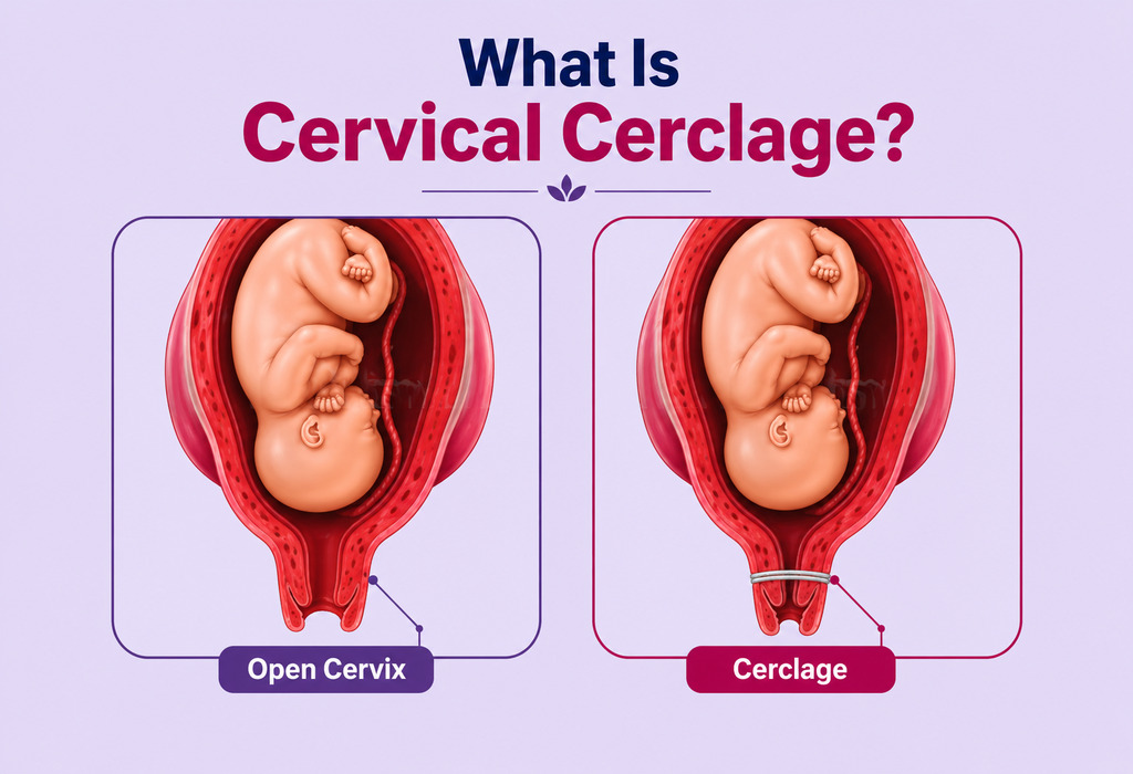
As your baby grows, the increasing weight can place pressure on a weakened cervix. Under the pressure, a weak cervix may start to widen during the second or third trimester, which can increase the risk of miscarriage or very premature delivery (preterm birth). Cervical cerclage in pregnancy is a known medical procedure that helps support the cervix when there is a higher risk of it opening too early during pregnancy. Cervical cerclage during pregnancy involves stitching the cervix closed to keep it closed for longer and improve the likelihood of carrying the pregnancy further. This procedure is typically performed before any signs of premature labour manifest, and is usually recommended for women with a history of cervical insufficiency or certain ultrasound findings.
Keep reading to find more about the different cervical cerclage methods, aftercare, risks, and success rate!
What Is Cervical Cerclage?
As per the Cleveland Clinic, cervical cerclage in pregnancy is a minor surgical procedure which is used to treat a weak cervix. By cervical cerclage meaning, we understand that in this procedure, the cervix is stitched closed to prevent miscarriage or premature delivery (1). The stitches are often removed in the last month of the pregnancy (2). This is mainly due to uterine cervical insufficiency, which is approximately seen in 0.5% of the general population and about 8% in women who have a history of second-trimester pregnancy loss (3).
While cervical cerclage lowers the chances of a miscarriage or pre-term labour, other problems related to early labour may arise after the procedure. Any woman who has had a cerclage should get herself examined routinely to avoid other complications.
Most women who require a cerclage in one pregnancy will need to have it placed in future pregnancies as well. This procedure has helped some high-risk pregnancies last longer, but it is not risk-free. The good news is that a successful procedure can lead to a full-term pregnancy (37 weeks).
Who Needs Cervical Cerclage and Why Is It Required?
Cerclage cannot be performed for women who have dilated more than 4 centimetres, who have ruptured membranes or in cases where the foetus has died. However, cervical cerclage is required in the following cases –
The mother-to-be has a history of late miscarriages because of an abnormal uterus or damaged cervix by abortion (4). A damaged cervix will not remain closed during pregnancy.
The mother-to-be had a previous 2nd-trimester pregnancy loss or a delivery that occurred with few or no contractions, suggesting that the cervix may not remain closed during pregnancy
Exposure of the foetus to diethylstilbestrol (DES)
The mother-to-be had any previous trauma or surgery of the uterus or cervix
Hormonal influences
Rupture of the membrane (water breaking)
The mother has a history of early births, and an ultrasound shows that your cervix is starting to open (5).
When Cervical Cerclage Is Not Recommended
Cervical cerclage is a procedure performed during pregnancy to reinforce the cervix, typically in cases where it is weak or at risk of premature dilation. However, cervical cerclage may not be recommended when:
The pregnant woman has vaginal bleeding
The pregnant woman is having uterine contractions
The membranes have ruptured early
The stitches have to be taken out prior to labour
The delivery requires a C-section to be done
What Are the Risks of Getting a Cervical Stitch?
Cervical cerclage is generally a safe procedure. However, there might be some complications arising after the procedure. Emergency cerclages have a higher risk of complications. The risks of getting a cervical stitch during pregnancy are:
Risks associated with anaesthesia
Premature labour
Rupture of membranes prematurely (water breaking)
Chances of infections in the cervix
Chances of the cervix getting torn (it occurs if the stitch is not removed at the start of labour)
Excessive bleeding
Chances of miscarriage increase in presence of cerclage
Infection of the amniotic sac
Cervical dystocia and failure to dilate requiring C-section
A displaced cervix
Foetal malformations in case of laparoscopic cerclage (6)
Types of Cervical Cerclage
Prior to getting the cerclage, your medical history will be reviewed, including an examination of your cervix and transvaginal ultrasound. Your doctor will determine the procedure best suited for your case and will pick one of three options
The three types of cervical cerclage techniques are (7):-
Transvaginal Cerclage (TVC) – TVC may be placed anytime from the 12th week onwards. The TVC is removed around the 37th week so that there are chances of normal delivery after cervical cerclage.
Transabdominal Cerclage (TAC) – TAC is a permanent procedure; hence, the delivery has to happen through C-section.
Transvaginal Cervico-isthmic Cerclage (TVCIC) – TVCIC can be removed for vaginal delivery or can be left if it is a C-section.
Benefits of a Cerclage
Cervical cerclage prevents miscarriage or preterm delivery or labour. According to the American Pregnancy Association, the success rate of this procedure is about 85% to 90% (4). This procedure is effective if there is truly a weak cervix; however, the diagnosis of this can be inaccurate.
How to Prepare for Cervical Cerclage Procedure?
Before the procedure is performed, a number of preparatory steps need to be completed –
A complete medical history will be taken and analysed.
A cervical examination will be conducted to assess the cervix.
Ultrasound of the vagina will be performed to check the baby’s vital signs and assess major birth defects.
Your doctor may also do an amniocentesis. Amniocentesis is the procedure in which a small sample of the amniotic fluid is retrieved and tested to identify any chromosomal abnormalities the foetus might have.
In case you have an infection that requires medication (antibiotics), you will need to complete treatment before the cerclage is done— if possible.
You will not be allowed any food or drink after midnight of the previous day from the surgery to avoid nausea and vomiting during and after the cerclage.
You will be advised not to have sexual intercourse, use tampons or douche for 24 hrs before the procedure.
An intravenous catheter will be placed to administer the fluids and medications.
The The Royal College of Obstetricians and Gynaecologists recommends that a cervical cerclage should be done at 11th to 14th week of pregnancy, when indicated by a patient’s history. When indicated via physical examination, the procedure can be done at up to 27 weeks, 6 days, and when indicated by ultrasound, it can be done at 14 to 24 weeks (8) (9).
Cervical Stitch Placement Procedure
In a cervical cerclage surgery, stitches are placed in the cervix, which is the outlet of the uterus, to hold it closed. This is carried out using anaesthesia (general or regional). An instrument is used to spread the vaginal walls, and the cervix is sewn in place. If you’re wondering about how long a cervical cerclage procedure takes, then the answer is about 30 minutes.
There are three ways in which the cervical stitches during pregnancy are done –
In this procedure, a stitch is placed through the vagina in and around the cervix like a purse string and placed at the bottom of the cervix as high as the doctor can reach. This is called Transvaginal Cerclage (TVC).
A band is placed around the outside of the cervix and then tied to control dilation. The band is placed either at the top or at the bottom of the uterus. It is placed laparoscopically or through an incision via the abdomen. In this procedure, the size of the cervix is not made smaller; the band stops it from dilating. This is called the Transabdominal Cerclage (TAC).
A small incision is made in the cervix, and a special tape is tied through it to close the cervix. This cerclage is placed above the cardinal ligaments but not as high as the TAC. This is the third type of cerclage, Transvaginal cervico-isthmic cerclage (TVCIC).
The cervical stitch may cause some pain, spotting or painful urination. Your doctor may prescribe Acetaminophen to help you get relief from the cervical stitch pain. If the doctor has placed stitches to reposition the vaginal tissues due to the incision in the cervix, you may notice the passage of the material in 2 to 3 weeks as the stitches take time to dissolve.
The cost of a cervical stitch varies depending on the medical clinic and doctor you are consulting.
Cervical Cerclage Aftercare
It is important that you recover well after the cervical stitches have been placed. Your doctor may keep you under observation for a few hours to ensure that you do not go into premature labour, post which you will be allowed to go home with restrictions and guidelines to be followed. The process of recovery after cervical cerclage includes medication, reduced or no physical activity, abstinence from sex, and regular weekly or bi-weekly visits to the doctor.
Constipation: After the cerclage has been placed, do not try to push the bowel movement too hard. You may want to make changes in your diet, like having high-fibre foods and more fluids. If required, you may use over-the-counter stool softener medicines too.
Rest: Ample rest will be required post the cerclage procedure.
Vaginal care: The vaginal area should be carefully washed with soap and water. You should avoid placing anything inside your vagina during this time.
Seek medical assistance immediately if –
You feel something bulging through the vagina
Clear fluid starts coming out of the vagina
If you have pain in the lower abdomen
Foul-smelling vaginal discharge starts coming from the vagina
If you experience regular contractions
If there is vaginal bleeding
If there is pain while urinating
Cervical Stitch Removal
The removal of cervical cerclage happens in or around the 37th week or at the onset of preterm labour.
In case of any emergency, it may be removed earlier.
Removing the ‘Transabdominal Cerclage (TAC)’ is a painless procedure which can be performed without any anaesthesia. It may cause light bleeding sometimes. You may go into labour (normal delivery) within 24 hours after the cervical stitch is removed.
If you have had a transabdominal cerclage, then you will be advised to go for a C-section birth and the stitch may be left in for any future pregnancies (10).
For ‘Transabdominal Cerclage (TAC)’ removal or the removal of a cervical stitch during pregnancy, surgery has to be performed. The cerclage may be left in place in case you plan to have a C-section.
What Are the Results?
In a woman who has had a previous preterm delivery and who is pregnant and has a short cervix (less than 25 mm), the result of cervical cerclage will be preventing a preterm delivery and reducing the chances of death and illness in the baby.
Cervical Cerclage in Twin Pregnancy
A cervical stitch is not always necessary for a twin pregnancy. However, your doctor may advise you to have this procedure if cervical length shortens significantly or if there is a history of preterm birth.
Alternatives to Cervical Cerclage
Depending on the condition, a woman may have some alternative therapies available to her to avoid or delay premature labour instead of cervical cerclage. These include:
Bed rest after cervical cerclage is prescribed so that there is less pressure on the cervix.
There are drugs that are designed to stop or delay labor (Ritrodrine, terbutaline, and magnesium sulfate)
Antibiotics may be prescribed to prevent preterm delivery.
Complications in Cervical Cerclage
Your doctor will explain all the risks and complications which may arise in the cervical cerclage procedure. If you experience any of the following symptoms after the stitch is placed, then you should consult the doctor:
Pain in abdomen or lower back
Vaginal bleeding
If you have fever of over 100 degrees
Nausea or vomiting
Foul smelling vaginal discharge
Water breaking or leaking
Success Rate of Cerclage Stitch
Performing a cerclage stitch of a combination of procedures cannot assure a successful full-term pregnancy. The doctors at the most reduce the risk for you and your baby. Cerclages work best when they are placed early in pregnancy. The success rate for cervical cerclages is between 85% and 90%. The success rate is calculated by assessing the number of pregnancies delivered closer to term with the procedure. Emergency cerclage has the lowest success rate.
Precautions to Take for Cervical Stitching During Pregnancy
Some precautions after a cervical stitch have been placed ensure the success of the procedure. The following precautions need to be taken if you have undergone cervical cerclage –
Complete bed rest if you have undergone cervical cerclage, depending upon your medical condition.
Reduced physical activity, along with no exercise, is also recommended. In case you want to exercise, ensure medical supervision and/or approval.
Refrain from heavy work for a short time.
Avoid sexual activity for some time (up to the 34th week) after the procedure.
It is advised to avoid climbing stairs after having a cervical procedure. However, there is no scientific evidence yet to prove that this could cause any issues. Since every case is different, you may seek advice from your doctor about what your specific requirements are.
How Will It Affect My Pregnancy?
A cervical cerclage during pregnancy may affect you in the following ways –
In most cases, a woman with a cervical cerclage will be able to complete the full term of pregnancy. The stitch is usually removed in the 37th week.
If you have undergone a transvaginal cerclage, you may go into labour within 24 hours of the stitch removal. You will not experience any pain.
If you have undergone a Transabdominal Cerclage, you will need to have a C-section.
In case you have to go in for premature labour and the drugs taken to delay the labour are not working, the stitch has to be removed as an emergency. This is done to avoid the risk of any damage to the cervix.
If your waters break early and contractions do not start, it is recommended to remove the stitch within 48 hours. The reason is a high risk of infection if it is delayed.
If you feel that you are going into labour before the stitch is removed, then you should immediately get admitted and inform the doctor. Leaving the stitch during the labour could lead to serious damage to the cervix.
Pregnancy After a Cerclage
There is no evidence that future pregnancies will be affected if you have had a cerclage before. However, in most cases, the doctor prefers putting a cerclage as a preventive measure if you have had one in your previous pregnancy.
10% of pregnancies end in preterm delivery (delivery before 37th week). Premature birth is a cause of serious health problems, like respiratory issues, problems regulating body temperature and infections. According to statistics, preterm delivery causes more than 85% of long-term disabilities and 75% of deaths among newborns.
When to Consult a Doctor?
It is essential to consult a doctor if you experience any signs that may indicate cervical insufficiency or complications during pregnancy. Some situations that warrant immediate medical attention include:
Unusual vaginal discharge or bleeding
A high fever
Regular uterine contractions or cramping
Severe pelvic pressure or discomfort
A sudden gush of fluid indicating ruptured membranes
History of second-trimester miscarriages or preterm births
Early consultation with a healthcare provider can help in timely diagnosis and management of potential issues, ensuring the best possible outcome for both mother and baby.
FAQs
1. Is cervical cerclage a painful procedure?
The procedure is done using a pain-numbing medication, such as a local anaesthetic. So, you won’t feel any pain or discomfort during the procedure. You may feel like a little pinch when the anaesthetic needle is inserted into the cervix to numb it. After the procedure, you should not feel much pain, but similar to the pain of an average menstrual period. It is also normal to experience some light bleeding or spotting and cramping a few days after the procedure.
2. How long is the cerclage left in?
The cervical cerclage procedure is performed between 12th and 14th weeks of pregnancy (at the beginning of the 2nd trimester) before symptoms of premature labor begin. Emergency cerclages are those placed at a later stage in pregnancy when cervical changes have already begun.
The cerclage can be left till the 37th week of pregnancy, after which it can be removed. If you plan to go in for a C-section, then the cerclage can be left inside. It can be removed before your water breaks or contractions start.
3. Is it possible to have a normal delivery after a cervical cerclage?
Yes, it is possible to have a normal vaginal delivery after a cervical cerclage. Typically, the stitches are removed around the 37th week of pregnancy or earlier if labour begins. After the stitches are removed, if there are no other complications, a normal vaginal delivery can be attempted.
4. Can cervical cerclage be performed more than once in subsequent pregnancies?
Yes, cervical cerclage can be performed in subsequent pregnancies if the patient had a successful outcome in a previous pregnancy with the procedure. The decision to repeat the cerclage will depend on the individual’s medical history and the current pregnancy’s circumstances.
5. What are the chances of infection after cervical cerclage?
While cervical cerclage is generally safe, there is a small risk of infection. Signs of infection may include fever, unusual discharge, or abdominal pain. It is crucial to follow your doctor’s post-procedure instructions and report any unusual symptoms promptly to minimise the risk of infection.
A weak cervix may pose a threat to the safety of your pregnancy. The Cervical Cerclage procedure will add a stitch of precaution to avoid any mishaps during the course of pregnancy.
Also Read:
Cervical Incompetence
Cervical Length during Pregnancy
Cervical Cancer in Pregnancy
Cervix & Cervical Position in Early Pregnancy
Cervix Dilation during Labour and Childbirth
Read more

Cholestasis of Pregnancy: Signs, Symptoms, and Treatment
Pregnancy is not easy for all expecting moms. There are several conditions that may affect a woman while she is pregnant and pose complications. One such condition is cholestasis. Cholestasis is a condition in which the liver cannot excrete the bile properly. This condition can cause severe itching to a pregnant woman and can pose complications for the baby.
What Is Cholestasis?
Also known as obstetric cholestasis, it is an uncommon condition that occurs during pregnancy and affects the liver, causing itching during pregnancy. This condition is seen in 1 in 70 pregnant Indian women. This condition causes the blockage of the normal flow of bile in the body. Medical practitioners now prefer to call it intrahepatic cholestasis of pregnancy or ICP.
Because the flow of bile is blocked, the bile builds up in the liver. The bile, particularly the bile salts, then begin to leak into the bloodstream. When this happens, it makes the skin extremely itchy, especially of the hands and feet. It usually manifests in the last trimester of the pregnancy. While this condition does not seem to have any long term effects on the mother’s health, it can cause severe complications for the infant.
What Are the Causes of Obstetric Cholestasis (OC)?
There aren’t any clear reasons as to why cholestasis of pregnancy occurs. However, there are hypotheses that hormonal and genetic factors might play a role.
1. Hormonal Factors
Some women might be highly sensitive to the changes in hormonal levels caused due to their pregnancy. The increase in oestrogen and progesterone affects the liver in a way that slows down the bile as it flows through the tiny ducts.
2. Genetic Factors
Women who have obstetric cholestasis in their family history have more chances of developing the condition themselves. This is because it seems to be inherited, though it is known to skip generations. These women may have inherited a problem in the way the bile is produced and moved through the body. While this problem does not manifest otherwise, a change in the hormone levels during pregnancy could cause the condition to flare up.
Other reasons for the development of this condition include disease, infection and certain medicines.
What Are the Symptoms of Obstetric Cholestasis in Pregnancy?
The following cholestasis liver disease symptoms may be present if you are suffering from OC during your pregnancy:
Itching: This is the primary symptom of the condition and often affects the palms of your hands and the soles of your feet. The itching because of cholestasis itching will be worse at night though there will be no rash.
Jaundice: This is a rare symptom of OC. Your eyes and skin might turn yellow which is an indication of jaundice.
Dark urine: Urine passed by a pregnant woman suffering from cholestasis will be dark in colour.
Light coloured stools: Stools will be lighter in colour than normal/regular stools.
Tiredness: Cholestasis can leave a pregnant woman feeling drained and out of energy.
Loss of appetite: Cholestasis affects hunger and eating schedule.
If you experience intense itching around your feet and palms, do talk to your doctor immediately. Obstetric cholestasis can cause severe complications to the health of your baby and if you have this condition, then you and your baby will be monitored closely.
Risk Factors for Obstetric Cholestasis
There are many factors that can increase the risk of the condition manifesting in pregnant women. These include:
If a woman has multiple pregnancies, she will have higher chances of developing OC.
Any previous history of liver disease or injury could increase the chances of the manifestation of OC.
Family history can be an indicator of the condition.
What Are the Complications of Cholestasis of Pregnancy?
Cholestasis of pregnancy affects both the mother and baby, though it affects the health of the baby more so. Here are some of the ways in which cholestasis of pregnancy can cause complications.
1. Complications for the Mother
The effects of cholestasis on the mother are usually non-life-threatening. The condition can temporarily affect how the body absorbs fat-soluble vitamins though this will hardly affect her nutrition. The itching usually resolves itself within a few days after delivery and the condition does not cause any long term effects on the liver.
2. Complications to the unborn baby
Obstetric cholestasis causes the baby to be born prematurely though the exact reason why is unknown. The foetus has a liquid known as meconium that could leak into the amniotic fluid. If the foetus then inhales the meconium during delivery, she will have complications with her breathing. The condition could also cause foetal demise in late pregnancy. Due to the high chances of stillbirth due to obstetric cholestasis, your doctor might want to induce labour by the 37th week.
Diagnosis and Test for Cholestasis in Pregnancy
Your doctor will suggest a blood test for detecting obstetric cholestasis called liver function test or LFT. Apart from this, she could also ask for a fasting serum bile acid test. If tests are negative, but the itching continues, they must be repeated.
Your doctor might also suggest an ultrasound to detect obstetric cholestasis. This could rule out gallbladder stones. Other tests to rule out abnormal liver functions like viral hepatitis, Epstein Barr virus, and cytomegalovirus are conducted before a diagnosis of OC can be made.
While all these tests may seem intimidating, you must remember that they are not complicated and are extremely useful for helping maintain the health of your baby.
What Are the Treatments for Obstetric Cholestasis?
Your doctor will recommend treatments that will reduce the itching and spread of the condition and avoid the risk of stillbirth. The treatments include the following methods:
Ursodeoxycholic acid is generally administered for the itching and to restore the liver back to normal functioning.
In cases of extreme cholestasis, the doctor will administer steroids.
The leaking bile salts will cause a loss of Vitamin K which is required for blood clotting. Thus, Vitamin K supplements can be prescribed.
As soon as the baby develops lungs, your doctor will be ready to induce labour to avoid stillbirth. This is usually between week 35 and 38.
Are There Any Natural Remedies for Obstetric Cholestasis?
Many women find that relaxing provides relief. You can use the following methods, particularly at night, so that you can fall asleep easier.
Lower the temperature of the room by cranking up the air-conditioning.
Do not use a blanket and stay uncovered while you sleep as heat can worsen the itching.
Take a cool shower or bath right before bedtime as cholestasis itching worsens at night due to circadian fluctuations in pregnancy hormones, and taking a bath with cold water can provide you temporary relief and help you sleep better.
Soak your hands and feet in ice water for a while to get relief from the itching
Some women have also found that using a mild moisturising lotion has provided them with some relief. Using a lotion with menthol in it has helped others. Before you do use any lotion, check with your doctor first.
Eat a healthy and balanced diet with minimal oily and fatty food to reduce the pressure on your liver.
Use only mild soaps so as to not further irritate your skin.
Wear loose clothing to let your skin breathe.
How Can You Prevent Potential Complications of Obstetric Cholestasis?
Due to the complications posed by obstetric cholestasis, your doctor might recommend close monitoring of your baby. These measures may include:
1. Non-stress Tests and Biophysical Profile Scores
Through this test, your medical practitioner will monitor how much your baby moves at certain times and how much her heart rate increases with the movement. The scores of the biophysical profile will give your doctor information about the volume of amniotic fluid as well as fetal muscle tone and activity.
2. Inducing Early Labor
Even if all the prenatal tests offer normal results, your doctor will recommend that she induce early labour by the 37th week. Since it is near impossible to predict when foetal death can occur, it is best to opt for early delivery.
FAQs
1. Will the mother get Obstetric Cholestasis in another pregnancy?
The chances of a pregnancy with obstetric cholestasis occurring after a previous pregnancy with the condition are very high. If the condition occurs in your family, you too could have a pregnancy with this condition.
2. Will my baby get delivered early if I have Obstetric Cholestasis?
Since the chances of stillbirth in this condition are very high, your doctor will recommend inducing labour between the 35th and 38th week. This is usually done around the 37th week.
3. What happens after labour & childbirth?
Your doctor will take the following measures to ensure that your health as well your little one’s health stays uncompromised after childbirth.
A vitamin K injection will be administered to your baby to ensure that the baby doesn’t bleed.
You will have a follow-up liver test after six to 12 weeks to check the health of your liver and rule out the presence of OC.
An ultrasound will be done to rule out gall stone formation.
You will not be given birth control pills that have estrogen in them. Talk to your doctor about other options for contraception.
Obstetric cholestasis can be an uncomfortable condition for you and can cause serious complications for your baby. Keep a note of all your symptoms and keep your medical practitioner in the loop. Maintain all appointments with her to ensure the safe delivery of your baby. Talk to your doctor about solutions for the itching and make sure you keep yourself stress-free to relieve discomfort.
Also Read:
Hives (Urticaria) when Pregnant
Stomach Itching in Pregnancy
Vaginal Itching while Pregnant
Jaundice in Pregnancy
Itchy Feet During Pregnancy
Read more
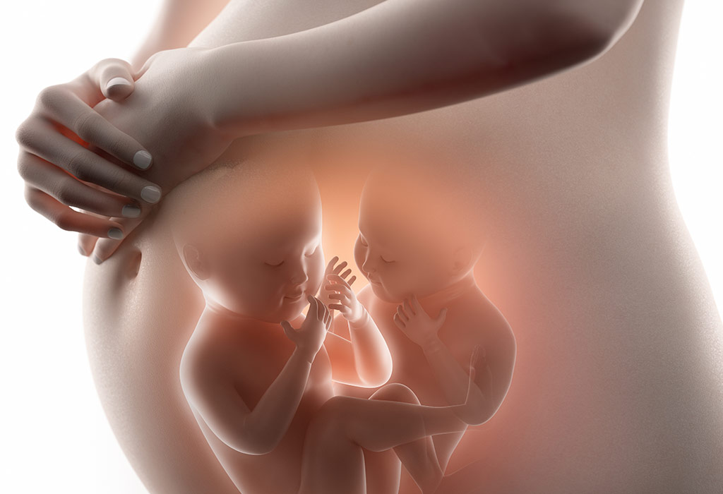
Twin Pregnancy Complications Mother and Baby
Twin pregnancies carry a higher risk of complications for both the mother and babies than singleton pregnancies, which is why they are classified as high-risk pregnancies and require closer medical monitoring. Research from the WHO Multicountry Survey found that 37.1% of twin pregnancies result in preterm birth, and twins are significantly more likely to experience low birth weight, NICU admission, and other adverse outcomes compared with singletons (1).
While many women with twin pregnancies deliver healthy babies, carrying two babies places extra demands on the mother’s body and increases the likelihood of complications. In this article, we’ll explore the most common complications of twin pregnancy, the risks they pose to both mother and babies, factors that increase the likelihood of complications with twins, and the medical strategies used to manage them effectively.
Why Twin and Multiple Pregnancies Are a Concern?
A multiple or a twin pregnancy may put your health at risk. The chances of developing gestational diabetes, preeclampsia, and other pregnancy-related complications are higher in the case of multiple pregnancies due to the greater load on the mother’s body and immune system with two or three babies (2).
Twin Pregnancy Complications
The chances of complications during twin pregnancy are higher when the twins share the same placenta. Dichorionic diamniotic twins complications (twins not sharing the same placenta) are not that severe as compared to monochorionic diamniotic twins complications (twins sharing the same placenta).
Here are some twin pregnancy risks you need to watch out for (3):
1. High Blood Pressure
A rise in the level of the blood pressure is 2-3 times more likely in you if you are expecting twins (or more babies) than you if you expecting one.
2. Preeclampsia
Preeclampsia is another risks of twin pregnancy that you might have to face. When you are expecting more than one baby, the strain on your placenta is more compared to a single baby (4).
If you are expecting twins, your doctor will closely monitor your blood pressure levels and test your urine to diagnose preeclampsia symptoms, if any, at antenatal appointments. The doctor might also prescribe aspirin-type drugs which will be started in the first trimester and might be continued later too to reduce your blood pressure levels and prevent preeclampsia. The screening is advised in the following cases:
If this is your first pregnancy
If you are above 40 years of age
If your BMI is 35 or more than that
If there is a history of preeclampsia in your family
If you became pregnant after a gap of 10 years
3. Gestational Diabetes
The risk of gestational diabetes is also higher with twin or multiple pregnancies. The doctor will check your urine to know your blood sugar levels. A glucose tolerance test will be prescribed if your doctor suspects that you have diabetes (5).
4. Anaemia
Your iron levels may drop due to an increased level of blood flow and the growing needs of the babies, leading to anaemia (6). You need to be extra careful about your iron levels as the drop will make you feel tired. Your doctor will recommend iron supplements. However, make sure you include vegetables that are rich in iron in your diet as well. Anaemia is more common in mothers expecting more than one baby. Your doctor will closely monitor you and also give your more blood tests if required.
5. Obstetric Cholestasis
This rare condition is caused by oestrogen and progesterone hormones and it affects your liver. When you are expecting twin (or more) babies, the secretion levels of these pregnancy hormones go up and lead to obstetric cholestasis. Symptoms include severe itching (7).
6. Placental Abruption
This is a serious condition that happens when your placenta detaches from the uterine wall before the delivery (8). This is one of the most serious twin pregnancy complications the third trimester poses.
7. Postpartum Hemorrhage
Women carrying twins are at a higher risk of postpartum hemorrhage, which refers to excessive bleeding after childbirth. This happens because the uterus becomes more stretched than usual when carrying two babies, making it harder for the uterine muscles to contract effectively after delivery (9).
Complications That Can Affect the Babies
Here are some of the complications which may affect the babies in a twin pregnancy.
1. Foetal Restriction
Retarded growth of the babies, where one or both the babies do not grow as much as they should and this can lead to complications both during birth and labour (10). However, this is something that you do not have to worry about much. Most twins are born small but they are healthy. Your doctor will conduct regular scans to monitor the growth of your twins and this would help in detecting all kinds of problems.
2. TTTS or Twin to Twin Transfusion Syndrome
This happens when one baby shares the other baby’s blood supply. The condition is rare but can lead to serious problems. This is a common complication among monochorionic diamniotic twins and it affects as much as 15% of the identical twins. One of the babies gets too much blood while the other gets much less than what is required. As a result, the baby donating blood is smaller and looks anaemic too due to the lack of blood. A higher volume of blood strains the heart of the other baby and this can lead to a heart failure (11).
3. Entangled Umbilical Cords
Monoamniotic Monochorionic twins are twins with entangled umbilical cords. This is a rare condition which prevents the flow of oxygen and nutrients to the developing babies (12).
4. Congenital Abnormalities
The risk of congenital abnormalities in the babies is more in the case of multiple pregnancies.
5. Premature Birth
Twins are more likely to be born prematurely than single babies. A premature birth occurs when babies are delivered before 37 weeks of pregnancy (13). Because twins often have less space to grow in the womb, labor may start earlier than expected or doctors may recommend an early delivery if complications arise.
What Are the Chances and Risks of Premature Delivery?
The risk of preterm labour and miscarriage is also higher and so are the chances and risks of premature delivery. However, it is hard to say how much higher this risk exactly is. This is because early miscarriages are hardly easily identified. If you do face a premature delivery, you can’t take your babies home immediately. The extent to which they need to stay in the hospital will depend on how premature they are.
In the cases of twins, full-term is 37 weeks. More than 50% of the twins are born before the 37th week and less than 10% are born before the 32nd week.
What Are the Chances of Having a Miscarriage?
The chance of miscarriage with twins are higher compared to when you are carrying a single child. It is common for miscarriages to happen during the initial twelve weeks, whether it is single or multiple pregnancies.
Chances and Risks of Stillbirth
The risks of stillbirth are higher in cases of multiple pregnancies. Losing the baby in the first month after the birth is also possible.
In case of multiple pregnancies, the risk of the complications including stillbirth increases just after the completion of the 38th week. This is the reason why doctors suggest induction or a caesarean if you do not deliver the twins by the 38th week of your pregnancy.
The rate of stillbirth is more in the case of identical twin babies who share the same placenta. The mother is often advised to deliver earlier, by the 37th week, in such cases.
Vanishing Twin Syndrome
In many cases, one baby is lost and if you lose the baby within the first trimester, the development of the other baby is usually not affected. The lost foetus is reabsorbed completely, a condition known as vanishing twin syndrome, and you might not experience any symptoms. The percentage range of this syndrome is 21-30 percent, meaning it is quite common (14).
Preventing Complications of Twin or Multiple Pregnancies
Your behaviour or lifestyle has very little to do as far as multiple or twin pregnancy is concerned. Getting the early confirmation that you are carrying twins or triplets will give your doctor the chance to prevent and treat all twin or triplets pregnancy risks that may arise.
Always stay hydrated and well-nourished. Never miss even a single prenatal appointment with your gynaecologists and make sure to abide by all the instructions given by your healthcare provider. Keep the following tips in mind to prevent any sort of complications.
Tips to Remember When Pregnant With Twins
The odds of conceiving twins cannot be determined exactly in any case. Keep the following tips in mind to keep yourself healthy with multiple pregnancies (15):
Maintaining a healthy weight is crucial in the case of twin pregnancies. The weight you can gain before 20 weeks is crucial. Your weight should be such that it reduces the risk of a low birth weight and premature birth.
Your iron levels are most likely to drop as the blood flow increases in the case of twin pregnancies. This can lead to anaemia and this will be harmful to both you and the babies. Take iron supplements regularly, after consulting your doctor.
Going for antenatal appointments is very important. Your doctor will conduct regular check-ups and ensure that you and your babies are healthy.
FAQs
1. Can one twin grow slower than the other?
Yes, one twin may grow more slowly than the other, a condition known as selective fetal growth restriction (sFGR). Regular ultrasounds help doctors monitor the babies’ growth and identify any concerns early.
2. Do all twins need to stay in the NICU after birth?
No, not all twins require neonatal intensive care. However, twins are more likely to need NICU support if they are born prematurely, have a low birth weight, or experience health complications after delivery.
Knowing the risks and complications of multiple pregnancies will go a long way in alleviating any concerns you may have about carrying multiples. Stay aware and do make sure you speak to your doctor if you are worried at any point in your pregnancies.
Also Read:
Types of Twins
Twins with IVF
Twin Pregnancy Precautions & Care
Read more

Molar Pregnancy (Hydatidiform Mole): Signs & Treatment
A molar pregnancy is a rare pregnancy complication in which abnormal tissue grows inside the uterus instead of a healthy embryo and placenta. It affects approximately 1 in every 1,000 pregnancies, making it uncommon but medically significant (1). It can occur in different forms, including a partial, complete or full molar pregnancy. In even rarer situations, a molar pregnancy twins case can occur, where a normal fetus develops alongside abnormal molar tissue.
If you’ve recently received a diagnosis following a 9 week molar pregnancy scan or are looking to understand this condition better, this article explains its causes, types, symptoms, diagnosis, treatment options, possible complications, recovery, and future pregnancy outlook.
What Is a Molar Pregnancy?
A molar pregnancy meaning is, after fertilisation, the embryo is abnormally developed and appears like a bunch of grapes, called a hydatidiform mole. It is a rare developmental disease of the cells that form the placenta, and is usually detected in the early trimesters and in most cases, results in wasted pregnancy (2).
Types of Molar Pregnancy
Based on the genetic and chromosomal pattern, molar pregnancies are divided into complete and partial molar pregnancy (3).
1. Complete Molar Pregnancy
During fertilisation, 23 chromosomes each are received from the mother and the father, and the fertilised egg hence contains 46 chromosomes altogether. In a complete molar pregnancy, the 23 maternal chromosomes are missing, and the embryo consists of only 23 chromosomes that are paternal in origin (4). This leads to the absence of foetal growth and formation of abnormal cysts, which appear like grapes.
2. Partial Molar Pregnancy
In a partial molar pregnancy, the embryo consists of 23 maternal chromosomes, but these chromosomes are fertilised by double the number of paternal chromosomes, 46 instead of 23. This leads to a total constitution of 69 chromosomes (4). The resultant embryo may grow and develop into an immature foetus, which may not survive beyond the few initial months of intrauterine growth due to the unfavourable genetic makeup.
What Are the Causes of Molar Pregnancy?
The reasons of molar pregnancy is largely unknown. However, the following factors have been associated with an increased risk for molar pregnancy (5):
Extremes of Age: It is more common in teen pregnancies and those after the age of 40.
Ethnic Origin: It is twice more common in women of Asian origin.
Past History: Women with a history of molar pregnancy in the past, are at an increased and proportionate risk. One such pregnancy in the past, predisposes to a risk of 1-1.5%, while two or more such past pregnancies increase the risk to 15-20% chances of having a molar pregnancy again.
Signs of Molar Pregnancy
A molar pregnancy may be asymptomatic or present with the usual symptoms of a normal pregnancy in the early few days. With advancing gestational age, it may present with the following symptoms (4):
Abnormal vaginal bleeding: first or early second trimester.
Disproportionately large uterus: large cysts may be present.
Metabolic and gastric symptoms: nausea and vomiting.
Autonomic instability: hypertension, sweating, palpitations, diarrhoea, etc.
Discomfort: dull pain in the lower pelvis.
Small cysts: Clusters of grape-like substance from the vagina usually suggest molar pregnancy. This symptom is specific for molar pregnancy.
However, these symptoms are highly non-specific (except the last one) as they may also occur in normal pregnancy or the case of a miscarriage.
Who Are More Likely to Have Molar Pregnancy?
A molar pregnancy can affect any woman of reproductive age, but certain factors are known to increase the risk. While having one or more of these risk factors does not guarantee that a molar pregnancy will occur, being aware of them can help with early monitoring and diagnosis (2) (5).
Women younger than 20 and those older than 35, especially over 40, are at a higher risk of experiencing a molar pregnancy.
Having a previous molar pregnancy significantly increases the chances of developing another molar pregnancy in the future.
A history of multiple miscarriages may be associated with a slightly increased risk of abnormal pregnancy development.
Nutritional deficiencies, particularly low levels of vitamin A (carotene), have been linked to a higher risk in some studies.
Women from certain geographic regions, including parts of Asia, tend to have higher rates of molar pregnancy than those in North America and Europe.
Rare inherited genetic abnormalities that affect fertilisation can increase the likelihood of recurrent molar pregnancies.
How Common Are Molar Pregnancies?
Worldwide, the condition is more common in women from the Asian subcontinent where it occurs at a frequency of 1 in every 1500 pregnancies. It is also more common in women who had a molar pregnancy in the past.
How Is the Diagnosis Made?
Diagnosis of molar pregnancy can be based on laboratory studies and imaging studies.
Beta HCG (human chorionic gonadotropin) levels: It is a hormone secreted by the placenta soon after fertilisation of the ovum during pregnancy. It can be measured in the blood as well as the urine. In a normal pregnancy, the levels range between hundreds (IU/ml) and proportionately increase with increasing gestational age (6). In molar pregnancies, the levels of B-HCG are enormously raised, exceeding 100000 IU/ml in few cases.
Molar pregnancy ultrasound: It is the investigation of choice for the diagnosis and confirmation of a molar pregnancy (7). An ultrasound shows a ‘snowstorm pattern’ with multiple cysts like growths in the uterine cavity and an absence of foetal activities. It is the safest, quickest and most reliable diagnostic tool for a molar pregnancy. It also confirms the diagnosis made by abnormally high beta HCG levels.
Risks and Complications of Molar Pregnancy
Molar pregnancies can be associated with the following risks and complications:
Invasive mole or choriocarcinoma: Complete molar pregnancies can progress to invasive mole or choriocarcinoma (8).
GTN or Gestational Trophoblastic Neoplasia: Molar pregnancies are associated with a risk of conversion to trophoblastic malignancy in the future. These are localized malignant conditions with a fairly good prognosis (9).
Recurrence: Women with a previous molar pregnancy have higher chances of a molar pregnancy recurrence compared to those with no prior history of the condition.
Foetal loss: Complete moles usually result in foetal loss. Women with molar pregnancies are at a high risk of miscarriages and other pregnancy-related complications as the genetic makeup of the foetus results in its premature death.
Molar Pregnancy Treatment
Treatment for a molar pregnancy focuses on removing the abnormal tissue from the uterus and preventing potential complications. The following treatment options may be used in the management of molar pregnancy:
Suction Removal or Dilation and Curettage(D&C): When a molar pregnancy is suspected by symptoms, hCG levels or ultrasound, a Dilation & Curettage is usually undertaken. It simply involves dilation of the cervical canal and suctioning the entire contents or spooning or curetting of the uterine contents to histopathologically confirm the diagnosis and therapeutically remove all the trophoblastic tissues (10).
HCG Monitoring: In a few cases after treatment, some residual tissues are left behind in the uterine cavity after a D&C. This is correlated with serial monitoring of beta HCG levels, which ideally should start falling after treatment. In majority cases, the residual tissues are successfully treated with However a fraction of cases progress to gestational trophoblastic neoplasia.
Medication: Medical treatment of molar pregnancy includes (5):
Symptomatic therapy: correction of anaemias, anti-thyroid drugs.
Antineoplastic drugs: drugs like methotrexate are useful in gestational trophoblastic neoplasia.
Folic acid: supplements with high amounts of folic acid.
Hysterectomy: For women with advanced age, or completed family or recurrent severe disease despite optimum medical therapy, hysterectomy is the best-suggested treatment option (11).
Monitoring After the Treatment
To avoid any residual disease, monitoring after treatment is essential. Regular follow-ups with your clinician after treatment, for serial physical examinations and history, are vital.
Beta hCG levels are the best means of monitoring after treatment with the help of serial monthly blood and urine tests (12). Normally, the very high molar pregnancy hCG levels should fall significantly after treatment. If it persistently remains high, residual or recurrent disease should be suspected. Screening ultrasound can be performed to rule out any possibility of trophoblastic disease after treatment.
Further Treatment After Molar Pregnancy
Despite curative treatment available for molar pregnancy, a few cases (1% partial and about 15% complete) may have a residual tissue, and this is known as a persistent trophoblastic disease (PTD). It may have malignant behaviour with spread to other body tissues, commonly involving the lungs. Treatment involves chemotherapy with methotrexate cycles along with folic acid supplementation.
Prevention of Molar Pregnancy
Since the pathophysiology is largely genetic, no definite preventive measures are available. However, following steps can be considered:
Balanced diet include adequate carotene and other green vegetables in your daily diet.
Avoid cigarette smoking and alcohol consumption.
Avoid conceiving for at least a year after previous molar pregnancy.
Consult a genetic counsellor and consider gene studies and chromosomal mapping.
Chances of Molar Pregnancy in Future
Molar pregnancies have a 1.5-2% risk of recurrence. No serious long-term health hazards are reported. One may have a normal pregnancy after a molar event. However, a gap of six months following surgery and about a year after chemotherapy is advisable before conceiving to avoid recurrence.
When Can You Try to Get Pregnant?
Fortunately, molar pregnancy or its treatment does not affect the fertility. Also, it has a low (1.5-2%) risk of recurrence. Clinicians worldwide advice a waiting period of 1 year before conceiving. This gives time for the beta hCG to return to the physiological levels. Once pre-pathological levels are recorded, you may now plan a safe pregnancy.
How to Cope With Fear and Loss?
A molar pregnancy can be a traumatising experience, and a grave loss of your child like in miscarriages. Strong will power and your partner’s support is vital. Undue anxiety and over treatments to avoid recurrence can be dangerous. Consulting a counsellor and considering adoption are advisable.
When to Consult a Doctor?
Early medical attention is essential if you experience symptoms that could indicate a molar pregnancy or another pregnancy-related complication. While some signs may seem similar to those of a normal pregnancy, unusual symptoms should never be ignored, especially during the first trimester.
Consult a doctor if you experience (2):
Vaginal bleeding during pregnancy, especially dark brown or bright red bleeding.
Severe nausea and vomiting that interferes with eating or drinking.
Rapid enlargement of the uterus or a belly that seems larger than expected for the stage of pregnancy.
Severe pelvic pain, pressure, or abdominal discomfort.
Passing grape-like cysts or tissue from the vagina.
Symptoms of high blood pressure before 20 weeks of pregnancy, such as severe headaches, blurred vision, or swelling.
FAQs
1. Is a molar pregnancy considered a miscarriage?
A molar pregnancy is not a typical miscarriage, although it cannot result in a viable baby. It is classified as a type of gestational trophoblastic disease and requires specific treatment to remove the abnormal tissue (2).
2. Is molar pregnancy bleeding always heavy?
Not necessarily. Molar pregnancy bleeding can range from light spotting to heavy vaginal bleeding. It often occurs during the first trimester and may appear dark brown or bright red in colour.
3. Can a molar pregnancy become cancerous?
In a small number of cases, molar tissue can persist after treatment and develop into gestational trophoblastic neoplasia (GTN). Regular follow-up monitoring helps detect and treat this condition early (7).
Molar pregnancy is a rare pathological condition that involves loss of pregnancy. With a good understanding of molar pregnancy meaning, timely evaluation, and complete treatment, this condition can be managed, and its recurrence prevented.
Also Read:
False Pregnancy
Ectopic Pregnancy
Chemical Pregnancy
Viable or Nonviable Pregnancy
Read more
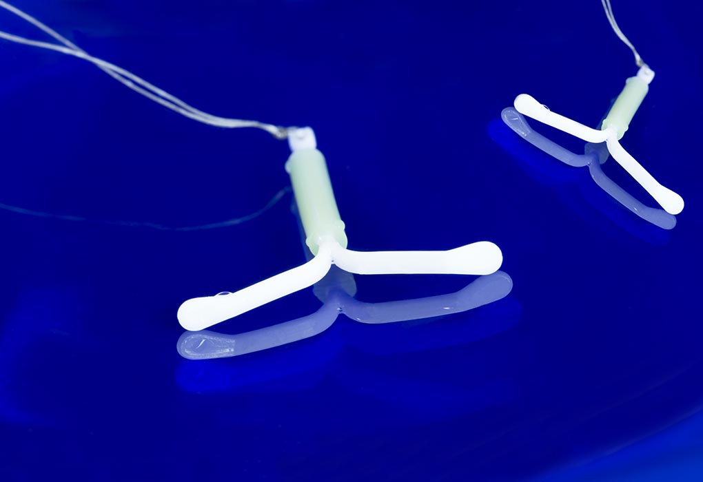
Pregnant With an IUD – Signs, Symptoms, and FAQ's
The intrauterine device (IUD) has become a favourite choice for many women because it’s simple, low-maintenance, and highly effective. Once placed in the uterus, it can work for 3 to 10 years, depending on the type used. With a success rate of more than 99%, it’s often considered one of the most dependable forms of birth control. But here’s the thing: no method is perfect. In rare cases, pregnancy can still occur with an IUD in place, and when it does, it may bring certain risks and complications for both mother and baby.
In this article, we’ll look at how pregnancy with an IUD happens, the risks involved, and what you should know if you ever face this situation, so keep reading to find out more.
Can You Get Pregnant With an IUD?
Although intrauterine devices are one of the most effective forms of birth control, their rate of being effective rate is about 98%. Therefore, there is a narrow margin, and it is observed that the odds of pregnancy with IUD are 3 cases out of 100. The American Congress of Obstetricians and Gynecologists (ACOG) says that the failure rate of IUDs during the first year of use is less than 1%, the same as sterilisation procedures like “tube tying” (1). In rare cases where pregnancies do occur, it’s usually caused by the device slipping partly or completely out of place. In fewer cases, the IUD may move and become embedded in the uterus and lose its effectiveness.
Signs & Symptoms of an IUD Pregnancy
Unlike the oral contraceptives, IUDs do not cause any changes in the body after it is put in place. Ovulation is not blocked, the endometrium continues to grow, and menstruation continues as normal (although, this depends on the type of IUD, which may cause bleeding to become much lighter or have it reduce altogether). The IUD only offers mechanical protection by not allowing the fertilised egg to implant in the uterine wall. In rare conditions, this protection can fail, and pregnancy proceeds as normal. Early pregnancy symptoms with the IUD is same as a normal pregnancy. These symptoms characterise it:
Delay in menstruation
Morning sickness and dizziness
Increase in the basal body temperature
Feeling tired, drowsy or fatigued
Lower abdominal pain
Loss of appetite in some women, conversely, excessive hunger
What to Do If You Suspect Pregnancy While Having an IUD?
It is not uncommon for women to worry about pregnancy when they are on an IUD. Many experience unusual bleeding for the first few months after the device is inserted. It is then normally followed by periods that are lighter and shorter; some even stop having periods entirely. Therefore, if you suspect that you are pregnant with IUD in place, follow these three steps (2):
1. Take a Pregnancy Test
If you feel symptoms of pregnancy and need to confirm it, the best way is to take a pregnancy test. A home pregnancy test is reliable. If you’re still in doubt, schedule a blood test for pregnancy with your doctor.
2. Visit Your Doctor
When you become pregnant, especially with an IUD in place, there’s a significant risk of an ectopic pregnancy (a pregnancy where the fertilised egg implants outside the uterus). Ectopic pregnancies are emergencies; therefore you need to visit your doctor immediately for a diagnosis.
3. Have the IUD Removed
If your pregnancy is viable and non-ectopic while the IUD is still in place, your doctor may recommend its removal (3). The doctor or a trained staff does this. It’s important not to try and remove it by yourself, as it is unsafe for the foetus.
Risks of IUD Pregnancy
IUDs are used to prevent pregnancies, so what happens if you get pregnant with an IUD? Knowing the risks of IUD and pregnancy can help you make better health decisions. Here are the main risks to be aware of (3) (4):
1. Miscarriage
A miscarriage is a significant risk when women get pregnant with an IUD. If the IUD is left in place during the pregnancy, the miscarriage rate goes up by 40-50 per cent. Removing it early on reduces the risk; however, it is still higher than normal women who got pregnant without an IUD (5).
2. Preterm Birth
Leaving the IUD in place during pregnancy also increases the chances of preterm birth along with miscarriage. It is seen that women who have an IUD in place during their pregnancy have five times more chances of giving birth prematurely than women who don’t. Removing the IUD early on lowers the risk of preterm birth.
3. Infection
Susceptibility to chorioamnionitis is high among pregnant women who have IUDs. It is an infection of the membranes surrounding the fetus and the amniotic fluid which can be potentially life-threatening (6). Removing the IUD early in pregnancy lowers the risk of the infection but does not remove it completely.
4. Placental Abruption
When the placenta separates from the uterine wall either before or during the delivery, it is called placental abruption. There could be a link between having the IUD in place and placental abruption (5).
5. Hormone Exposure
Hormonal IUDs pose a risk to the foetus as they slowly release progestin into the uterus. The long-term effects on babies are not fully understood. However, increased risk of congenital disabilities does not appear to be among the risks.
Is There an Instance Where a Doctor May Suggest ‘No IUD Removal’?
If you wish to continue your pregnancy, ideally the IUD should be removed. It is done if the strings are visible from the cervix and can be grasped. The pregnancy will then continue without complications, although there are slightly higher chances of miscarriage, infections and preterm labour. When the risk of removing the IUD outweighs the risk of leaving it in place, your doctor would suggest a ‘No IUD Removal’. There are instances where women have given birth to healthy babies even with the IUD. It usually so happens that as the baby grows; the IUD gets pushed out, somewhere around the middle of the term.
Possible Reasons for IUD Failure
Even though IUDs are one of the most effective birth control options available, they aren’t completely fail-proof. Here are some common reasons why an IUD might not work as intended:
Expulsion of the IUD: Sometimes the IUD can partially or completely slip out of the uterus, especially within the first few months of insertion. This reduces its effectiveness and may go unnoticed.
Improper Placement: If the IUD isn’t inserted correctly or shifts from its intended position, it may not provide full protection against pregnancy.
Using an Expired IUD: Each IUD has a lifespan of 3 to 10 years depending on the type. Using it beyond this timeframe increases the chances of failure.
Type of IUD and Timing of Insertion: Copper IUDs, such as Paragard, provide immediate protection once inserted. However, hormonal IUDs, like Mirena, can take up to 7 days to become fully effective.
Tips to Protect Yourself From an Unwanted IUD Pregnancy
When used perfectly, IUDs are very effective at preventing pregnancies. Here are some steps to make sure that an unwanted pregnancy doesn’t occur with the IUD.
1. Visit Your Gynaecologist Regularly
Once a year, visit your gynaecologist and have an ultrasound scan of your uterus done. It can reveal any displacement of the IUD or even its loss. These two are the major causes of unwanted pregnancy when using an IUD.
2. Monitor the IUD Strings
Once a month, you can check if the device is properly in place by feeling for the strings attached to the device. They extend down from the cervix to the top of the vagina and can be felt with your fingers. An increase in their length would mean that the device has been displaced into the cervix. The disappearance of the strings mostly indicates a loss of the IUD. Both of these conditions mandate a visit to the gynaecologist and a pregnancy test if the device is missing.
3. Know the Expiration Date
All IUDs have an expiration date; 3-5 years on an average after which they must be removed or replaced with a new one. When left beyond their time, they may embed themselves into the uterus walls making it difficult to remove and increase the chances of infections.
When to Consult the Doctor
If you have an IUD, it’s important to reach out to your doctor if you:
Are planning to become pregnant or suspect you might be pregnant.
Think your IUD may have moved or slipped out of place.
Want to have your IUD removed or replaced.
You should also contact your doctor if you experience any unusual symptoms while using an IUD, such as:
Fever, chills, or other signs of infection.
Severe or persistent lower abdominal pain or cramps.
Unusual vaginal discharge or heavier than normal bleeding.
Pain or bleeding during sexual activity.
Most side effects of an IUD are minor and temporary, but in rare cases, serious complications can occur. These may include ectopic pregnancy, bacterial infection, or perforation of the uterus, which require immediate medical attention (3) (4).
FAQs
1. Does age affect the risk of IUD failure?
IUD effectiveness is generally high across all ages, but younger women may have slightly higher expulsion rates due to a more active uterus.
2. Can heavy periods affect IUD effectiveness?
Heavy menstrual bleeding doesn’t usually reduce effectiveness, but in some cases, copper IUDs can worsen bleeding, which may lead to removal or monitoring (4) (7).
Many women have questions about the odds of getting pregnant with an IUD. With proper usage, IUDs are very effective at preventing pregnancy. In the rare event that you do get pregnant with one in place, consult your doctor immediately.
Also Read:
Pregnancy Symptoms after IUI
Get Pregnant with Irregular Periods
Is It Possible to Get Pregnant after IUD Removal?
Read more
Preeclampsia in Pregnancy
Preeclampsia is a disease that affects pregnant women and has been known to cause issues such as poor liver function and fluid in the lungs. Apart from affecting the mother, it also has the potential to cause debilitating complications for the child such as cerebral palsy, blindness and deafness due to preterm birth.
What Is Preeclampsia?
Formerly called Toxemia Pregnancy, preeclampsia is a pregnancy complication that can start as early as 20 weeks of pregnancy, in the second trimester, or anytime up to six weeks post-delivery, and can lead to issues such as kidney damage and high blood pressure. Mothers-to-be may not display any symptoms if they are afflicted by preeclampsia, which is why most doctors insist on checking on your blood pressure during every visit to the clinic. If the blood pressure is high, they will suggest a urine test to check the protein levels in it.
Once detected, it needs immediate treatment as it could lead to other health problems like eclampsia and HELLP syndrome, where HELLP stands for Hemolysis, Elevated Liver enzymes, and Low Platelet count.
Causes of Preeclampsia
A reduced blood flow towards the placenta is one of the primary causes of preeclampsia, and there are no visible symptoms. This happens when the placenta does not place itself properly in the uterus lining, and the dilation of the arteries in that area is not enough. Diabetes and chronic hypertension prior to pregnancy could also be reasons for less blood flow to the placenta.
It has also been found that when there is a variation in the blood flow to the placenta, certain placental proteins are released into the bloodstream in large volumes. Because of this, the following reactions are triggered off in your body:
The blood vessel walls are damaged, which leads to swelling of the body and an increase in the level of protein in the urine.
Blood vessels constrict, thus leading to high blood pressure.
Preeclampsia can be also be caused due to other factors such as auto-immune disorders, genetic factors, diet and issues related to the blood vessels. Your immune system and its reaction to the pregnancy is also thought to be one of the causes of preeclampsia.
Signs and Symptoms of Preeclampsia
Preeclampsia may or may not display any noticeable symptoms, and even when it does, the symptoms may differ from one woman to another. People often get confused as preeclampsia has symptoms similar to the normal symptoms of pregnancy such as nausea, weight gain, and swelling. The early stages of preeclampsia may not be typically noticeable and thus need careful monitoring during visits to the doctor.
Speak to your doctor if you notice any unusual swelling in any part of your body as it could be related to preeclampsia swelling. Watch out for the following:
Swelling of hands and fingers.
Excessive swelling of feet and ankles.
Swelling of the face and neck or puffiness around the eyes.
Rapid increase of weight in a short period of a week or a fortnight.
It is to be noted that every pregnant woman who gains rapid weight or has swelling may not be affected by preeclampsia, and the causes for her condition could be different.
Many pregnant women who are affected by preeclampsia have also experienced symptoms like headaches that do not seem to subside, and altered vision. Your doctor may also advise urine and blood tests to check the protein level in urine, platelet level and any abnormality of liver enzymes.
Abdominal pain, slower reflexes, less or no urine, nausea and vomiting, and dizziness are also common symptoms associated with preeclampsia. You should ensure that your blood pressure remains in the range of 140/90 and any increase or decrease of these levels warrants a quick visit to the doctor. Regular checks of blood and urine should keep you updated on this.
The following signs are warning signs that could signal preeclampsia:
Difficulty in breathing.
Vomiting or nausea (especially when it happens in the 2nd or 3rd trimester).
Temporary vision loss, extreme light sensitivity, double vision or blurriness.
Pain in the upper abdomen.
Who Is at the Risk of Getting Preeclampsia?
Those who have had preeclampsia during their first pregnancy are more likely to develop it during subsequent pregnancies. The risk of developing preeclampsia is higher depending on the severity of the condition and when it appears during pregnancy. This could mean that if you have contracted preeclampsia prior to 29 weeks of your pregnancy, then there is a 40% higher chance of contracting it again in later pregnancy.
Though scientific evidence is lacking, Preeclampsia has a high incidence rate among pregnant teens. One study suggests that lack of prenatal care from unwanted pregnancies leaves pregnant teens vulnerable.
Preeclampsia is high among pregnant women above 40 as it puts excess strain on their bodies and may kickstart underlying medical conditions.
Generally, obese women with a Body Mass Index (BMI) of over 30 have an increased risk of preeclampsia as obesity aggravates blood pressure.
Diagnosis of Preeclampsia
During your scheduled appointments with the doctor, your blood pressure and urine will be under immense scrutiny. Elevated levels of blood pressure and protein in urine are likely to show the presence of preeclampsia. To be diagnosed with preeclampsia, you will need to have:
1. BP more than 140/90 or diastolic more than 90.
2. Proteinuria
Doctors are now aware that elevated blood pressure is a must for pregnant women to be diagnosed with preeclampsia.
If your doctor suspects preeclampsia, the following tests may be required:
1. Protein in Urine
Your doctor will ask for a urine test that can detect the presence of protein in your urine sample. If this initial test is positive, you will be advised to collect urine after 24 hours so that it can be sent for testing. This test is known as the most dependable and accurate test for preeclampsia. The presence of 300 mg and above of protein in the urine is a sure sign of preeclampsia.
2. Monitoring of Blood Pressure
If your systolic reading is higher than 140 or the diastolic reading is higher than 90, your blood pressure is higher than normal. Since blood pressure fluctuates as per the time of the day, your doctor will ask you to check it at different times to confirm that it is high. This is also a reliable pointer towards the diagnosis of preeclampsia for a pregnant woman.
3. Protein-Creatinine Ratio
Creatinine is a waste product of the body that is filtered out by the kidney along with other wastes. The Protein-Creatinine ratio is a urine test that checks the presence of this waste product, and hence the doctor can understand if the kidney functioning is normal. This test needs one random sample, which is better than collecting the 24-hour urine for the urine test. If your test shows the presence of 0.3 mg/dl, it can be concluded that you have been afflicted by preeclampsia.
4. Foetal Ultrasound
This test is generally recommended to monitor your baby’s growth closely using the ultrasound method. In this manner, the doctor can estimate the foetal weight and gauge the level of amniotic fluid in the uterus.
5. Non-stress Test
This test involves a simple procedure that helps to check the baby’s heart rate and its reaction to movement.
6. Biophysical Profile
In this test, an ultrasound is conducted to measure the foetal breathing, movement, muscle tone and the amniotic fluid volume in the mother’s uterus.
Complications of Preeclampsia
Complications of preeclampsia are rare, but they can quickly develop into life-threatening problems, such as a low platelet count and a breakdown of red blood cells. Regular monitoring and quick diagnosis can ensure the complications do not grow, and the condition is arrested in time.
Let’s take a look at the complications this medical condition could possibly lead to in the mother and the child if it is left untreated.
1. Complications For the Mother
The following problems can affect the mother if she has been diagnosed with preeclampsia:
Eclampsia: This involves an involuntary contraction of the muscles and can be described as a fit or a convulsion that pregnant women can have. It can occur after the 20th week or immediately after delivery. During the fit, which lasts for less than a minute, the mother can experience repetitive movements of the arms, legs or neck, and she could also lose consciousness.
Stroke: When the blood supply to the brain is reduced due to high blood pressure, it can cause cerebral haemorrhage, commonly known as a stroke. In this case, the brain does not receive the necessary oxygen from the blood, causing the cells to die, and thus leading to brain damage or in certain cases, death.
Blood clotting: Medically known as Disseminated Intravascular Coagulation. In this condition, the mother’s blood clotting capacity is severely affected. Here, there is heavy bleeding since the protein level in the blood has drastically fallen, or there are a number of blood clots as the proteins have become highly active.
2. Complications For the Baby
If the mother is diagnosed with preeclampsia, the baby can face the following health issues:
Smaller Size of the Baby: Since there is a low supply of oxygen and nutrients to the baby during preeclampsia, such babies will be smaller in size. This is especially true if preeclampsia occurs prior to 37 weeks of pregnancy.
Breathing Difficulties in the Baby: In case the preeclampsia is diagnosed as severe, the doctor may take the decision to deliver the baby earlier. This can lead to breathing difficulties in the baby since the lungs are not fully developed.
Stillborn Baby: In some cases, the baby could be stillborn due to early delivery.
Earlier in this article, we spoke a little bit about HELLP syndrome; let’s take a look at it, too.
What Is HELLP Syndrome?
The HELLP syndrome is a rare blood clotting and liver disorder that is a severe version of pregnancy eclampsia. It is most likely to strike post-delivery, but cases have also been seen after 20 weeks, and sometimes, before 20 weeks, too. The acronym HELLP stands for each of the conditions:
H is for Haemolysis during which time the red blood cells in the bloodstream break down.
EL stands for Elevated Liver enzymes which is a sign of damage to the liver.
LP is for Low Platelet count which is responsible for clotting of blood.
Once the diagnosis is made, treatment should be given promptly for preeclampsia. Read on to know more.
Treatment for Preeclampsia When Pregnant
If you are 37 weeks pregnant or more, labour will be induced, especially when the cervix has dilated well. The doctor may also opt for a C-section if they feel that you or your baby will be unable to withstand the pressures of a normal delivery.
For the treatment of Severe Preeclampsia, you could be asked to remain at the hospital for regular monitoring if you are diagnosed with severe preeclampsia. A preeclampsia specialist could be assigned to you to give you special care and help in the management of the condition. Medication will be administered to lower the blood pressure along with an intravenous infusion of magnesium sulphate to help prevent seizures.
What If Preeclampsia Develops During Labour or After Labour?
If you are diagnosed with preeclampsia during or after labour, monitoring your condition will be of high priority. You will have to stay back at the hospital for a few more days to avoid any further complications that could develop if your blood pressure rises or you get seizures. You will be given magnesium sulphate for up to 24 hours after the delivery to avoid seizures. If you go home, you will have to report back with blood pressure checks conducted for at least a week.
Effects of Preeclampsia on Future Pregnancy
Preeclampsia or toxaemia in pregnancy is a serious condition. However, the mother continues to live with its effects, and the risk remains. The effects of preeclampsia on your organs may take at least six weeks to get resolved after delivery. Studies have also shown that there is also an increased risk of hypertension, Type 2 Diabetes, stroke and heart disease. The toxaemia in pregnancy affects the baby by limiting the supply of nutrients to the baby, which can change his structure and metabolism. This could lead to coronary heart disease and related disorders, including diabetes, stroke and hypertension.
How to Prevent Preeclampsia
Getting prenatal care and not missing on any of your appointments is the key to prevention of preeclampsia. You will have to keep a close watch on your blood pressure and the protein content in your urine to keep toxaemia health-related issues away. As soon as the first signs of preeclampsia are visible, alert your doctor so treatment can be started without any delay. Depending on the severity of the condition, the number of weeks and the baby’s condition, your doctor will decide the line of treatment. This will include numerous urine tests and blood pressure monitoring.
Preeclampsia is one of the foremost causes of maternal mortality and causes foetal mortality as well. However, close monitoring of health conditions of women with a history of high blood pressure and hypertension can be helpful in managing preeclampsia.
Also Read: Cramping During Pregnancy
Read more
Complications in the First Trimester of Pregnancy
It is often said that pregnancy is one of the most beautiful phases in a woman’s life. It involves pampering, indulgence, and immense support from one’s partner and family. Most pregnancies progress smoothly and without complications. However, understanding the possible complications of first trimester pregnancy is essential for every expectant mother. During the early stages of pregnancy, women undergo several tests, ultrasounds, and physical examinations so that doctors can rule out any complications or address concerns promptly.
An expectant mother is more susceptible to certain risks as the body undergoes tremendous changes to accommodate new life. Certain parts of the immune system become more active, while others are naturally suppressed to support these changes. Here is a look at some common early pregnancy problems and solutions every expecting mother should know about.
Pregnancy Complications in the First Trimester
There is a significant change in hormonal activity during the first trimester. This change is essential but brings with it complications like fatigue or nausea. While these are low risk and are more bothersome than dangerous, there are a few serious pregnancy problems in first trimester that you should be aware of. We have mentioned them below:
1. Miscarriage
It is unfortunate that about 10-15 per cent of pregnancies conclude in a miscarriage. Of these, it is reported that around 80% happen during the first trimester. Miscarriage during early pregnancy can be caused by infections, chromosomal abnormalities, clotting disorders, or anatomic problems with the uterus. Eating a balanced diet, appropriate exercise, avoiding alcohol and cigarettes should help sustain your pregnancy (1).
2. Ectopic Pregnancy
An ectopic pregnancy happens when a fertilised egg gets implanted outside the uterus. One in 50 of all pregnancies could be an ectopic one. Most ectopic pregnancies occur in the fallopian tube and may cause the tube to rupture. This can be fatal as it causes internal bleeding (2).
3. Constipation and Diarrhoea
Stomach problems during pregnancy first trimester are not uncommon. Usually, women experience mild to severe constipation due to changes in diet and incorporation of iron-rich foods. This may sometimes cause haemorrhoids and bloody stools (3). Diarrhoea that lasts more than 3-4 days should be reported to the doctor, or it could lead to dehydration and weakness. Continue to have a balanced diet and drink copious amounts of water. In rare cases, diarrhoea can be a result of food poisoning. It should be treated immediately if it doesn’t resolve within 24 hours (4).
4. Multiple and Twin Pregnancy Complications
30% of mums carrying twins or multiple foetuses are known to have vaginal bleeding. Twin pregnancy complications in the first trimester include vanishing twin, where one baby is lost to a miscarriage during the pregnancy. The embryo that is lost is usually reabsorbed into the uterine lining without any noticeable symptoms to the expectant mother (5). Up to 30 per cent of multiple pregnancies face the vanishing twin syndrome. It is detected because the mother has usually had early scans that showed dual embryos.
5. Vaginal Discharges or Itching
Vaginal discharge is a regularity in a woman’s life. Nothing is alarming about a little vaginal discharge unless it has an odour or is coloured yellowish. Additionally, if there is itching or redness, it could indicate an infection. A more serious concern is if the itching indicates an STD that could be transmitted to the baby (6).
6. Painful Urination
A urinary tract infection is a bane in every woman’s life. During pregnancy, a woman becomes highly susceptible to Urinary Tract Infection. The changes that pregnancy causes to the urinary tract makes it very easy for a pregnant woman to contract a UTI. Additionally, the pressure from the uterus prevents the bladder from completely draining and this causes infection. If you leave a UTI untreated, it can lead to kidney infection or early labour (7).
7. Difficulty Breathing
Increased levels of progesterone during the first trimester contribute to shortness of breath. Working slowly, sitting up straight, and sleeping on propped up pillows might help with easing breathing problems during the first trimester of pregnancy. Once you’re past the first trimester, the shortness of breath usually occurs around the third trimester when your uterus pushes up against the diaphragm and lungs, making it difficult to take a deep breath. If wheezing, palpitations, or coughing accompany shortness of breath, then you should let your doctor know immediately. They may be able to tell the difference between plain shortness of breath and other complications.
8. Spotting or Vaginal Bleeding
Implantation bleeding is common in pregnancy and is nothing to get alarmed about (8). There are, however, other reasons like cervical polyps that can lead to spotting and bleeding (9). Rupture of a tubal pregnancy, as in the case of an ectopic pregnancy, can also cause a fatal bleed.
9. Calf Pain & Headache
Calf pains during the first trimester are not very common. Hormonal changes combined with exhaustion can be credited with this. But pain on one side alone could be an indicator of a travelling blood clot. Pregnancy increases the chances of developing clots. A clot in the head could cause severe headaches. Clots that form in leg veins are known as Deep Vein Thrombosis. Staying active and improving blood circulation reduces the chances of blood clots and cramps (10).
10. Nausea & Vomiting
Morning sickness is the most common affliction that affects a pregnant woman during her first trimester. It affects different women differently. For some people, it could be extreme, and it increases the risk of dehydration. Some women cannot hold down anything that they eat. It may lead to reduced nutrition for the mother and foetus. Doctors may recommend some medication to regulate nausea and prevent dehydration in extreme cases (4).
11. Fatigue & Insomnia
Pregnant women, during the first trimester, experience a massive slump in the quality of sleep. Pregnancy sleep problems in the first trimester could be attributed to the fatigue and extreme exhaustion that women feel throughout the day. It could also be attributed to heartburn and bathroom breaks at night.
12. Temperature Above 101°F
A fever that crosses 101°F could be a sign of an underlying infection. This could invariably affect the foetus, and it should be checked. Fever accompanied by rash or joint pain could be parvovirus or Toxoplasma (11). There is also a minor chance that it could indicate cytomegalovirus which is a reason for congenital deafness (12). The chances of risk to the infant occur when a pregnant woman has recurrent CMV infection.
13. Swelling
After 20 weeks of pregnancy, swelling is considered to be normal in most women. This is caused by a change in blood volume and blood flow, allowing fluids to be trapped in the tissues around the extremities. But swelling in the first trimester, especially in the face and hands could be an indicator of preeclampsia (13). Always consult your doctor if in doubt about such things.
FAQs
1. How can women reduce the risk of early pregnancy problems?
Women can lower the risk of complications by eating a balanced diet, staying hydrated, taking prenatal vitamins, avoiding smoking and alcohol, managing stress, and attending regular prenatal checkups. Early medical guidance plays a key role in ensuring a healthy pregnancy for both mother and baby.
2. What are the signs of an unhealthy pregnancy in the first trimester?
Certain symptoms such as severe abdominal pain, persistent vomiting, high fever, heavy bleeding, dizziness, or sudden loss of pregnancy symptoms may indicate a problem during early pregnancy. While some discomfort is normal, any unusual or severe symptom should be discussed with a doctor immediately.
Pregnant women might face any number of discomforts ranging from slight dizziness to mild pelvic pain. Once past the 12 weeks mark, discomforts like nausea and vomiting will reduce or completely stop. Expectant mothers will now be asked to get scans and tests done in each trimester to rule out any other complication like Down’s syndrome in the foetus, preeclampsia, placenta previa, gestational diabetes, high blood pressure, or low amniotic fluid.
Bear in mind that although we have listed one too many 1st trimester pregnancy problems that mothers-to-be should watch out for, pregnancy is one of the most naturally beautiful phases in a woman’s life. There are many positives about being pregnant like hearing baby’s first heartbeat, feeling that first movement, the first kick, the glow and radiance of being pregnant, and the arrival of those tiny fingers and toes after 10 months. So, be watchful and heed any medical advice from your doctor but remember to relax and enjoy these months before your entire life becomes about your tiny tot.
Also Read:
Common Second Trimester Complications
Uterine Prolapse During Pregnancy
List of Pregnancy Problems and Their Solutions
Pregnancy Complications Every Woman Should Know
Read more
MTHFR in Pregnancy: Risks, Diagnosis & Treatment
Pregnancy can be a tough time for some women, especially for those who have tested positive for a mutated MTHFR gene. All humans have the MTHFR gene in their bodies, it is known as gene 5-methyltetrahydrofolate and is responsible for breaking folic-acid to create folate. Less folate or malfunctioning of the MTHFR gene can result in some disorders or health conditions, more so in pregnancy. In case the gene is mutated, it can increase the chances of miscarriage, preeclampsia, or birth defects in babies.
What Is Meant By MTHFR?
MTHFR or gene 5-methyltetrahydrofolate is a gene which is responsible for breaking down of folic acid to folate. It is found in every human and is the cause of how the body processes folate. Folate in everyone is responsible for making their DNA, repairing the DNA and producing RBCs (Red Blood cells) in the body. In some people MTHFR gene malfunctions and stops or slows down the breaking of folic acid. This condition is known as mutated MTHFR gene. Having a mutated MTHFR is not an uncommon condition and is generally harmless in most people; however, in a few people, it can create certain health issues. People with positive MTHFR mutation gene have Hyperhomocysteinemia.
Hyperhomocysteinemia is the condition in the human body when the homocysteine levels are elevated. In women, high homocysteine levels with low folic acid levels may create complications in reproduction and lead to MTHFR mutation pregnancy. Women with mutated MTHFR gene may face issues in reproduction and may have a higher risk of preeclampsia, baby born with a certain birth defect such as spina bifida or miscarriage. Most females who have a problem in conceiving or repeated miscarriages or stillbirth come to know that they have mutated MTHFR gene. In some cases, females with mutated MTHFR gene can have successful pregnancies and may come to know of this condition much later or after suffering from a loss of their baby.
Risk Factors of Positive Mutated MTHFR
A mutated positive MTHFR gene is a genetic issue. It passes from the parents (mother or father) to the child. No one can have positive mutated MTHFR after birth, as a baby is born with it. Simply nothing causes positive MTHFR gene, it was always there. A person who has genetically inherited the positive mutated MTHFR from both parents may have more issues than a person who inherited it from one parent. However, having positive mutated MTHFR doesn’t mean one will have any health problem or issues. It just increases the risk of having health issues. A few of the risk factors of positive mutated MTHFR are:
Homocystinuria– It is a disorder where the body is able to process homocysteine at a lesser pace. This may result in eye problems, skeletal abnormalities, abnormal blood clotting, and cognitive problems.
Spina bifida– It is the incomplete formation or development of bones surrounding the spinal cord.
Anencephaly-It is a birth defect which is characterised by incompletely or missing formed parts in the skull or brain.
Hearing loss due to age.
Heart disease
Hypertension
Stroke
Preeclampsia or high blood pressure during pregnancy.
Psychiatric disorders
Glaucoma
Certain types of cancer
What Are the Complications of Positive MTHFR During Pregnancy?
MTHFR gene can have many different mutations. These mutations may cause various health problems and affect various organs such as the heart. Some of the mutations affect pregnancy more than the other mutations. Though there is no accurate scientific evidence which proves a positive mutated MTHFR gene can cause recurrent miscarriage; however, women who had suffered multiple miscarriages often test positive for mutated MTHFR gene. A positive mutated MTHFR gene may several complications during pregnancy. A few of the complications caused by positive MTHFR during pregnancy are:
1. Spina Bifida
Spina bifida is a birth defect in a baby where the spinal cord is sticking out of the back. This creates nerve damage. Several children can have normal lives if the severity of the Spinal Bifida is low. Babies with high severity need full-time care.
2. Anencephaly
Anencephaly is a serious birth defect. Babies suffering from anencephaly are born with an underdeveloped brain or skull i.e. some parts of their brain or skull are missing. Babies suffering from this defect mostly don’t live past one week of their birth.
3. Preeclampsia
Preeclampsia is a health condition of high blood pressure in pregnant women.
4. Blood Clotting
A positive MTHFR mutation can cause blood clotting during pregnancy between the uterine wall and growing placenta. This area is known as ‘low flow maternal-placental interface’ and the blood clot prevents the nutrients to be transported to the baby. MTHFR blood clotting disorder can occur both during early or late in pregnancy. When occurring in early pregnancy, it can cause repeated miscarriage as the foetus is very vulnerable. When it occurs in advance pregnancy, it causes stillbirth as the blood clot is formed in the umbilical cord or placenta.
Diagnosis of a Mutated MTHFR
A positive mutated MTHFR cannot be (and is) not tested in every pregnant woman. The test is expensive and the mutated MTHFR affects only a few women, so it is done only on the advice of the doctor. The doctor generally orders a test to detect mutated MTHFR in pregnant women who have had multiple miscarriages or had a family history of positive mutated MTHFR gene or the women who had genetic issues in their other pregnancies. The test done to determine positive mutated MTHFR involves the following testing methods:
The doctor may ask to check different variants of the MTHFR gene to check for positive MTHFR mutation through a blood test. The blood test is done to determine the homocysteine levels of a person.
Genetic testing is also done to identify the MTHFR gene mutation.
There are two gene variants which are most commonly tested. They are – C677T and A1298C.
A person having elevated homocysteine levels will have two C677T genes variant or one A1298C and one C6771 variant.
Any person with elevated homocysteine levels will not have two A1298C gene variants. Though it is possible to have high homocysteine levels with a negative MTHFR gene mutation test.
Treatment for Positive Mutated MTHFR
There are many treatments prescribed by doctors to treat positive mutated MTHFR gene. Most treatments help prevent blood clots and increase folic acid levels in the body. Some popular treatments for positive mutated MTHFR are:
1. Lovenox or Heparin Injections
These injections assist in preventing blood clots formation between the uterine wall and the developing placenta. Most doctors prescribe this treatment for pregnant women in their early pregnancy. This is because then the mutated MTHFR gene is not able to form a blood clot anytime during pregnancy. The doctor can only determine for how long the injections need to be continued.
2. Aspirin
Many pregnant women having mutated MTHFR gene often ask their doctors if they should take aspirin for MTHFR as Aspirin also works as a blood thinner. Aspirin may prevent blood clotting but it should be taken only after checking with a doctor.
3. Prenatal Vitamin with L-Methyl Folate
Anaemia is common in pregnant women. Pregnant women having mutated MTHFR gene generally have low folic acid. So MTHFR and folic acid are co-related. L- methyl folate helps in decreasing the anaemia risk in pregnant women. MTHFR prenatal vitamins are different than normal prenatal vitamins as pregnant women with mutated MTHFR gene process folic differently, so they need different prenatal vitamins with L-methyl folate rather than synthetic folic acid. So prenatal vitamins with L-methyl folate are prescribed instead of folic acids by some doctors.
Is MTHFR Linked to Miscarriage?
There are several opinions about MTHFR and pregnancy. Some people believe that positive MTHFR gene and miscarriages are directly linked and some believe there is no correlation. Though the increased number of miscarriages is often linked with positive MTHFR gene mutation as the women had C677T MTHFR gene. It is proved that women having C677T mutation cannot effectively recycle homocysteine which results in the accumulation of blood. Accumulation of blood results in homocysteinemia which poses a risk for coronary artery disease or formation of blood clots. Blood clots prevent nutrients transportation to the placenta, causing starvation of the foetus and eventually spontaneous abortion.
MTHFR gene mutation may seem scary especially for pregnant women. However, it can be managed with medications and proper treatment. Moreover, many females with positive MTHFR gene have had normal pregnancies. So there is no reason to panic. In case of any issues, talk to your doctor and she can determine the effects of MTHFR on your pregnancy. Timely diagnosis and prompt treatment can increase your chances of a successful pregnancy.
Also Read:
Placenta In Pregnancy
Eclampsia While Pregnant
Ectopic Pregnancy
Read more

HIV or AIDS in pregnancy: Symptoms, Risks, & Treatment
Pregnancy is often a time filled with excitement, hope, and careful planning for your baby’s future. But for women living with HIV, pregnancy often comes with additional questions: Will my baby be safe? Can I have a healthy pregnancy? What steps can I take to reduce the risk of transmission? Fortunately, medical advances have transformed the outlook for mothers and babies, making it possible for many women with HIV to give birth to healthy, HIV-negative children.
Want to learn about the effects of HIV during pregnancy, the treatments available, and the best ways to protect your baby? Read the full article to find out all about HIV/AIDS and pregnancy.
What Is HIV/AIDS?
HIV (Human immunodeficiency virus) is an infamous virus known to cause AIDS (Acquired immunodeficiency syndrome). As the name suggests, it’s a virus that gradually breaks down the immune system making you prone to opportunistic infections and various other complications. One point of confusion amongst people is that they believe HIV and AIDS are similar when that is not the case. A person may be HIV infected for years without having AIDS. You are HIV positive from the moment the virus is detected in your blood (1). But only after the symptoms set in and the immune system breaks down the person is said to have AIDS. It usually takes several years to reach the last stage of HIV – that is AIDS (2).
How Does HIV Spread During Pregnancy?
Unlike viral flu, HIV does not spread by contact, breathing the same air or through food and water. HIV can be spread in ways similar to the Hepatitis B virus (3):
Sexual intercourse (most common)
Blood / blood-contaminated products / other body fluid / organ transplantation
Vertical transmission: From mother to child via the placenta or breast milk
The chance of transmission depends on the viral load, that is the number of viruses per ml of the blood. Also, during pregnancy, high levels of the hormone progesterone increase the level of virus receptors. This aids the entry of virus and increases the chance of transmission.
Symptoms of HIV & AIDS
Once the HIV virus gets into the blood, it begins to multiply and infect a specific type of white blood cell called the T lymphocytes. It usually takes 3 to 6 weeks for the symptoms to set in which last for less than 10 days. These symptoms include (4):
Fever and night sweats
Fatigue
Rash
Headache
Swelling in neck, armpits, groin
Sore throat
Body ache, joint pains
Nausea, vomiting, and diarrhoea
Thus, the symptoms of an HIV positive pregnancy are not different from flu during pregnancy. Later, after the symptoms are gone, the virus continues to multiply and attack the immune system silently, until it breaks down. It may take as long as 10 years for this to happen.
Various Problems That AIDS Can Cause
In cases where HIV progresses to AIDS, the risk of health complications increases for both the mother and the baby. To assess immune health, doctors often check the CD4 count; a type of white blood cell that plays a vital role in fighting infections (5). A low CD4 count is a key indicator of advanced HIV infection or AIDS. Some of the possible complications associated with AIDS during pregnancy include:
Infections: As the CD4 count falls, more serious and deadly infections set in, tuberculosis being the most common.
Cancer: Various forms of cancer are common in AIDS. Women may suffer from genital tumours which may be cancerous.
STDs: Other sexually transmitted infections in addition to HIV like syphilis may add to the problem for the mother and child.
Factors Which Increase the Risks Associated With Transmission of HIV
Some of the issues that increase the likelihood of the spreading of AIDS include:
Viral load: The most important factor that determines the transmission is the viral load in the mother (6). For example, the rate of transmission is 1% if the load of the HIV virus in the maternal blood is less than 400 copies/mL, but this increases drastically to more than 30% when maternal viral levels are more than 100,000 copies / mL. But viral load in the blood may differ from that in the genital secretions. So, the transmission through genital secretions may occur even before it is detectable in maternal blood in some cases.
Pre-term delivery: There is a four-fold increased risk of a baby being exposed to the HIV virus in pre-term delivery.
Breastfeeding: If you are breastfeeding your baby, there is a 30-40% chance that your baby will be infected by the virus (7).
Mode of transmission: The mode by which the mother acquired HIV also determines the rate of transmission. If it was a sexually transmitted infection, the rate of vertical transmission to the baby is higher.
Initiation of anti-HIV treatment: The period of gestation at which the anti-HIV treatment was initiated to the mother affects the transmission to the baby.
Medical intervention: Some medical procedures done during delivery like forceps application, artificial rupture of membranes, and invasive fetal monitoring increase the risk of transmitting the virus from the mother to the baby (7).
Should Pregnant Women Get Tested for HIV?
It is mandatory for all pregnant women to undergo screening HIV test in some countries (opt-in approach) whereas in others the mother has the right to refuse after being counselled and informed about HIV (opt-out approach). Pregnant women who are injectable drug users, sex workers along with those who have HIV-infected sexual partners, multiple sexual partners or are diagnosed with an STD are recommended to repeat the test in the last trimester.
How Is HIV Testing Done?
HIV screening test is performed using an ELISA test (enzyme-linked immunoassay) (8).
If the test comes positive, it is confirmed with either a Western blot or Immunofluorescence assay (IFA) (9). A rapid detection using PCR can also be done.
Effects of HIV on the Mother’s and Baby’s Health
If the CD4 count is maintained high and the viral load is kept low it does not grossly affect the outcome of your pregnancy. All said and done, it is multifactorial and can manifest with variations. The real problems arise when HIV turns into full-blown AIDS.
1. Effect on Mother’s Health
The mother is at a high risk of various infections that can be life-threatening. Various cancers, both benign and malignant are common in AIDS which may affect the course of pregnancy. Some pregnancy-related complications like pre-term labour, hypertension, diabetes are common in HIV positive cases.
2. Effect on Baby’s Health
An HIV positive mother can also infect the baby. The infections acquired by the mother can be transmitted to the baby which can be life-threatening. It can potentially affect all the bodily functions of the baby (10).
Accuracy of HIV Test Result
HIV tests are highly accurate. The ELISA test has a sensitivity of more than 99.5%. This means that in more than 99.5 of 100 tests performed will pick up HIV if it is there. There is less than a 0.5% chance of missing HIV by ELISA. For further confirmation, the IFA and western blot have very high specificity. This means these tests very rarely will be a false-positive.
How Is HIV Transmitted From the Mother to the Child During Pregnancy?
The transmission that occurs from mother to child in the perinatal period is called vertical transmission. This transmission can be via the placenta and via breast milk.
1. Transmission in Early Pregnancy
Normally placenta forms a barrier between the mother’s and the baby’s blood. This forms a protective barrier for HIV transmission. But in very early pregnancy, while the fertilised egg is attaching to the uterus and the placenta is being formed, there is a chance of contact between their blood. Thus, HIV transmission can occur even before pregnancy is detected.
2. HIV and Labour: Transmission During Late Pregnancy
There is a high chance of transmission of HIV from mother to baby during the time of onset of labour. When the water bag ruptures, during the delivery process, and during placenta separation (10).
3. Transmission During Breastfeeding
It has been proven that the HIV virus is present in breast milk and babies who are breastfed are at an increased risk of being HIV positive.
Preventing Mother to Child Transmission of HIV & Its Challenges
Antiretroviral therapy (ART) is the anti-HIV medicines used as a remedial measure. ART plays a major role in preventing mother to child transmission of HIV (11). Being HIV positive and pregnant has a lot of challenges such as social stigma and non-medical hospital staff being hesitant to approach the patient. Many women suffer from anxiety in revealing that they have HIV may make them outcasts.
Here are some preventive measures which you need to keep in mind if you are trying to get pregnant (12):
Planning pregnancy: If you are planning to become pregnant, get yourself tested for HIV. If you test positive start right away with the antiretroviral therapy. This will not only reduce the viral load in the mother but also reduce the risk of transmission.
Post-exposure prophylaxis: If you are HIV negative but your partner has been diagnosed with HIV, you need to take the ART to prevent yourself from getting HIV.
Plan delivery: Planned c-section at 38 weeks is the preferred mode of delivery to minimise the risk of transmission of HIV.
Post-exposure prophylaxis for the baby: After birth, your baby will be given ART medicines to kill HIV virus if any has entered the baby’s blood from the mother.
Avoid breastfeeding: If good alternatives are available without compromising the baby’s nutrition, it is advised to avoid breastfeeding the baby.
With all these precautions taken the risk of transmission may be reduced to less than 1%.
Complications of HIV
HIV infection can add to the pregnancy-associated complications. These include (12):
Pre-term delivery
Low birth weight
Growth restriction of the baby
High blood pressure
Gestational diabetes
HIV Treatment
HIV treatment consists of antiretroviral therapy (ART). It consists of various regimens with different combinations of medicines either to be taken orally or injected. The regimen will be decided by your doctor based on the viral load, the CD4 count, previous treatments taken, the drug resistance pattern and the week of gestation. If you were on HIV treatment before conceiving, the treatment is usually continued.
Should Pregnant Women With HIV Take HIV Medicines?
The treatment of women infected with HIV should not be stopped because of pregnancy. Certain modifications in the regimen may be necessary as it may affect the development of the baby but the treatment needs to be continued throughout the pregnancy (10).
How Does HIV Affect Labor & Birth?
Usually, a C-section is advised at 38 weeks, but there is a high rate of preterm delivery and the associated complications in HIV infected mothers.
Some mothers may be given an injection of Zidovudine during labour which reduces the chance of transmission of HIV to the baby.
Certain procedures like artificial rupture of membranes to induce labour, using fetal scalp electrodes for fetal heart rate monitoring and delivery with vacuum or forceps are generally avoided given the potential of increased risk of transmission.
What If the Baby Tests HIV Positive?
All babies who test positive for HIV need to be given ART medicines. If the baby cannot tolerate oral medicines, then injectable medicines are given. It is a life-long treatment for the child. As with adults, there are different treatment regimens available for babies who test positive for HIV.
HIV Positive & Prenatal Care
As a protocol in hospitals, you will be treated like any other mother. Your healthcare professional will maintain professional secrecy. From counselling to appropriate treatment the healthcare professionals will take the necessary prenatal care required. As a pregnant woman with AIDS, the prenatal care responsibility that you have is to provide the doctor with all the information without maintaining any secrets.
Questions to Ask Your Doctor
If you have AIDS and you are pregnant, maintaining open communication with your doctor is essential for protecting your health and your baby’s well-being. Here are some important questions to discuss with your doctor:
1. How can I reduce the risk of transmitting HIV to my baby during pregnancy, labour, and delivery?
2. What medications will I need to take during pregnancy, and are they safe for my baby?
3. How often should my viral load and CD4 count be monitored throughout pregnancy?
4. Will HIV affect my pregnancy or increase the risk of complications?
5. What type of delivery is recommended for me based on my HIV status and viral load?
6. Can I breastfeed my baby, or should I consider alternative feeding options?
7. What follow-up care and testing will my baby need after birth to ensure they remain HIV-free?
FAQs
1. Does caesarean delivery reduce the risk of mother to child transmission of HIV?
There is only a 50% chance of the baby contracting the HIV virus if they are delivered via caesarean birth. In addition, the risk of transmission reduces by 87% when a caesarean section is combined with anti-HIV treatment.
2. Will my baby need treatment after birth?
Yes. Babies born to HIV positive mothers are given HIV treatment for 4-6 weeks after birth. This reduces the HIV multiplication if any, and protects the child.
3. Can an HIV positive mother breastfeed?
Breastfeeding is generally not recommended for mothers who are HIV positive as it significantly increases the risk of HIV transmission to the baby.
You can live a normal life with HIV. This includes becoming pregnant as well. Though there are complications involved, there is nothing stopping HIV positive women from getting pregnant. But you need to understand that despite all the precautions you may deliver HIV positive baby. As HIV reduces your immunity take extra precautions to keep yourself away from infections. Good hygiene, healthy food and exercise are key to a healthy pregnancy with HIV. With the advancements in HIV treatment, many babies born to HIV positive mothers test HIV negative.
Also Read:
CMV while Pregnant
Infections during Pregnancy
Trichomoniasis in Pregnancy
Sexually Transmitted Diseases in Pregnancy
Read more

Dilation & Evacuation (D and E) Surgical Procedure
Most women experience zero complications during pregnancy, and they go on to deliver and raise healthy babies. But for some women, things don’t go as expected, and they may have to end their pregnancies. It can be heartbreaking for a couple to make the difficult decision of ending their pregnancy, or it may result in a miscarriage. If a woman’s pregnancy needs to be terminated within the first thirteen weeks, a procedure called Dilation and Curettage is performed. However, if the pregnancy is further along, then Dilation and Evacuation (D&E) will be performed. In this article, we will explore everything about the D and E procedure, including how long it takes to complete, risks, complications, and more.
What Is Dilation and Evacuation?
Dilation and evacuation is a medical procedure that is performed in the second trimester of pregnancy to remove the remains of a fetus from the uterus. It combines dilation, vacuum aspiration and curettage. During D&E, suction and uterine forceps are used to aid in removing the remains of the foetus (1). While a D&E is usually done before the 15th week of pregnancy, women can also have a D&E at 20 weeks (2).
When Is D&E Abortion Done?
Women who need to end their pregnancies from the 13th week onwards can undergo the D&E procedure of abortion (3).
To Whom Is D&E Recommended?
There are various reasons why a woman may need to undergo a D&E procedure. A woman may be recommended D & E in the following cases:
When the baby dies in the womb. The baby will be stillborn, and the woman will be advised to have its remains removed, as it can sometimes lead to an infection.
If the baby is detected with extremely serious abnormalities, an abortion will be suggested.
If there is a premature rupture of the membranes, an immediate termination of the pregnancy may be prescribed.
In some cases, the woman’s health may be seriously compromised if she continues with the pregnancy. In such cases, she may be advised to terminate the pregnancy.
How Long Does It Take for the D & E Procedure?
The actual D&E (Dilation and Evacuation) procedure usually takes around 10 to 30 minutes, depending on the stage of pregnancy and the patient’s condition. However, the complete process may take longer because cervical preparation is often done several hours or even a day before the procedure (4).
After the procedure, the patient is typically monitored for a few hours before being discharged. Most women return home on the same day (5).
Tests Before The D&E Procedure
Before performing a D&E procedure, doctors may recommend several tests to ensure safety and reduce complications. These commonly include:
Ultrasound scan to confirm gestational age
Blood tests to check blood group and haemoglobin levels
Blood pressure and general health evaluation
Screening for infections if required
Review of medical history and current medications
These tests help the healthcare provider plan the safest treatment approach for the patient.
What Happens Before D&E?
Your doctor may write you an ultrasound to confirm the gestational age. Before the procedure, the cervix needs to be gently widened or softened to make the process safer. Doctors may use medications or special dilatants for cervical preparation.
The patient may also receive:
Medicine to soften and widen the cervix (given the night before the procedure)
Sedation or anaesthesia so that the patient doesn’t feel pain
Instructions about eating or drinking before the procedure
Antibiotics to lower infection risk
Counselling about recovery and aftercare
Healthcare providers usually explain the risks, possible side effects, and warning signs to watch for after the procedure. Emotional support and follow-up care are also important parts of the process.
Dilation and Evacuation Procedure Steps
Having to make the decision to terminate a pregnancy is very difficult for expectant couples. It can be very frightening for a woman to undergo such an experience, and not knowing what to expect can be helpful. Here is what to expect from D&E procedures (6):
Step 1: During the Surgery
Dilatants for abortion at this stage are necessary. The cervix is prepared around one or two days before the actual procedure is to be performed. This often requires the use of either seaweed or hydrogel rods. These absorb the moisture from the cervix and then expand it.
Another method that can be used to expand the cervix is through medication, but this is not preferred unless it is earlier in the 13th week.
Antibiotics are administered to prevent infection. General anaesthesia is administered before anything is done.
Once the anaesthesia has set in, the foetus and the placenta are removed from the uterus through the use of suctioning, curettage and forceps.
Step 2: After the Surgery
You will need to rest to allow your body to heal properly.
You will be put on antibiotics and other medications that help with shrinking the uterus.
Spotting or bleeding for two weeks after the procedure is normal. Use pads for this and avoid anything that needs to be inserted into the vagina. This is all to prevent abstaining from sex at this time.
Cramping is to be expected. The uterus will shrink back to its original size. This cramping may last for a few days, or it could be only for several hours. It depends on the individual.
Risks and Complications
While most procedures go off without a hitch, there are some occasions when you might face a few complications that will require serious medical attention.
Risks Associated With D&E Surgery
Here are some of the risks that are associated with the D&E procedure (7):
Injury can be caused in the lining of the uterus or the cervix.
Although rare, a hole can form in the lining of the uterus. This is called a uterine perforation. It usually forms during the dilation stage.
The uterus may not contract back into its non-pregnant size
Tissue can sometimes remain in the uterus.
Complications That Can Occur Later
Sometimes complications associated with the D&E procedure can arise at a later date. These complications include:
If you have developed a body infection as a result, you may experience muscle aches, headaches and dizziness. You could also go through a general feeling of unease or illness. Not all infections result in a fever as a symptom.
Severe bleeding, different from your regular menstrual bleeding, can occur. This bleeding can be excessive, and you may have to go through at least two large pads in an hour or less.
Fever or hot flashes that last for over four hours.
Swelling of the abdomen.
Vomiting that continues for over four hours.
Increased heart rate.
Swelling, pain or redness occurring in or around the genitals.
Increased vaginal discharge or discharge with a bad odour.
D&E Abortion – Cost
Dilation and evacuation abortion is an expensive procedure and can cost from around $500 to $3000 or sometimes, much more.
Taking Care After Undergoing a D&E Procedure
Having an abortion so late in the pregnancy can be very difficult for a woman. But if it is inevitable, you will have to go through it. There are a few things you should keep in mind to take care of yourself after undergoing a D&E procedure for your speedy recovery:
Remember that, as you have just gone through a very difficult procedure related to your reproduction, you will need to abstain from having sexual intercourse until your doctor says it is all right. Even when you get a go-ahead from your doctor, it is advisable that you ask your spouse to use a condom to lower the chances of infections. Birth control is another thing you will need to consider. Getting pregnant again so soon will not allow your body to heal as well as it should after going through something so traumatic.
For those who have had to undergo a D&E procedure, it is best that they visit a counsellor if they are unable to recover emotionally. You can even visit your therapist if you already have one. It is very important to speak to a professional, as there could be many factors that can lead to depression. Depression can also be a result of the changes in pregnancy hormones. Some of the most notable symptoms of depression are a change in your appetite or sleep, fatigue, constant sorrow, anxiety, emptiness and irritability. If these persist for more than two weeks, you should definitely seek counselling.
After the procedure, you will be kept under observation for about an hour or so. Once the observation period is over, your nurse will give you all the instructions you need to follow to recover physically.
Whether you undergo a D&E procedure due to a complication or a miscarriage, it can take a toll on your health. Your physical and mental health will suffer, but it is important that you speak to your doctor and open up to your loved ones to heal completely.
When to Consult a Doctor?
If you experience any of the following symptoms, it is advised to consult a doctor immediately:
Stomach pain that only gets worse
Swelling of your belly
Fluid from the vagina that increases or smells bad
Fast heart rate
Vomiting
Fever of 100.4°F (38°C) or higher
Feeling achy or unwell
New weird symptoms that get worse
Signs of depression that last for 2 weeks or more
FAQs
1. Is the D&E procedure painful?
The procedure is usually done under sedation or anaesthesia, so most women feel little to no pain during it. Mild cramping and bleeding afterwards are common.
2. How safe is a D&E abortion in the second trimester?
D&E is considered one of the safest and most commonly used methods for second-trimester abortion when performed by an experienced healthcare provider.
3. Will a D&E procedure affect future fertility?
In most cases, a properly performed D&E does not affect future fertility or the ability to get pregnant again.
4. How long does recovery take after a D&E procedure?
Many women recover physically within a few days, though light bleeding and cramps may continue for one to two weeks.
5. Is hospital admission required for a D&E procedure?
Most D&E procedures are done as outpatient procedures, meaning the patient can usually go home the same day unless complications arise.
Don’t keep your feelings bottled up. Speak to your gynaecologist about what you can do for a speedy recovery. Remember, undergoing a D&E procedure does not mean that you won’t be able to have a baby again. The D&E procedure does not affect a woman’s fertility. So give yourself enough time to heal; once you’re ready to have a baby, you can always try for another child if there are no health risks!
Also Read:
Nonstress Test
Medical Abortion
Surgical Abortion
Chances of Pregnancy after Abortion
Read more
Home Remedies UTI During Pregnancy – Natural Relief Tips
A urinary tract infection can be treated with home remedies for UTI during pregnancy, but where to start and where to look is a question among many pregnant women. A woman’s urinary tract can be a breeding ground for bacteria, more so during pregnancy. Since pregnant women are at an increased risk of UTIs between the 6th and 24th week of pregnancy, it is imperative you know what to do if you face one. Doctors usually suggest a urinalysis or urine culture to detect UTI during pregnancy (1). If the test results are positive, obviously you will worry because it requires immediate treatment or else it might damage the kidneys. Consulting a doctor is a must, but you can also try some home remedial measures to get rid of this infection. There are some natural remedies apart from the conventional treatment which can help prevent and treat UTI. But before we discuss home remedy for urine infection during pregnancy, let’s acquaint you with its symptoms.
Symptoms of UTI While Pregnant
If you have a UTI, you may experience one or more of the following symptoms, and you must inform your doctor about the same (1).
Blood or mucus in the urine
Burning sensation while urinating
Abdominal pain
Fever and incontinence
Cloudy or foul-smelling urine
The urge to urinate frequently
Natural Remedies for a UTI During Pregnancy
Most urinary tract infections, especially lower urinary tract infections, often subside on their own. But when they don’t, antibiotics are prescribed as the first line of treatment. However, not all antibiotics are safe during pregnancy. Trying home remedies for UTI when pregnant can help speed the healing process. Here are some home remedies for UTI in pregnancy!
1. Increase Your Fluid Intake
Drinking plenty of water and fluids daily can help flush out bacteria from your urinary tract by making you pee more and also prevent infection. To keep your body hydrated, drink water throughout the day and whenever you feel thirsty. This way, the bacteria will be flushed out of your system, lessening the chances of an infection, and help you treat a UTI while pregnant (2).
2. Urinate Frequently
During pregnancy, you must urinate often so that bacteria do not grow inside the bladder. If a person holds back urine for a long time, bacteria can multiply in the urinary tract, leading to an infection. But if you urinate frequently, it will put pressure on the bacteria and help clear them out from your bladder. So pee often and prevent and treat UTIs.
3. Take Probiotics
Probiotics support the human body’s flora which is used as a defence for the body. They promote a healthy balance of bacteria in the gut (keeping it free from harmful bacteria). Fermented foods like kimchi, probiotic yoghurt, and raw cheese are some of the healthiest probiotic foods. Eating these foods can help restore your body’s bacteria content without causing any disease (3).
4. Drink Cranberry Juice
The Cleveland Clinic suggests drinking unsweetened cranberry juice is one of the best remedies for UTIs. Cranberries help prevent UTIs from entering the urinary tract and thus prevent infection. Cranberry juice can also help prevent the growth of bacteria that might cause infection (4).
5. Use Clove Oil
Clove oil is known for its antimicrobial, antiviral, and antifungal properties. The antibacterial properties of clove oil help kill E.coli and prevent the chances of UTI. Topical application of clove oil can help prevent urogenital infections. However, using clove oil can also result in some adverse reactions on the body. Hence, it is suggested that you use it only after consulting with a doctor and under the supervision of your health care provider.
6. Increase Your Vitamin C Intake
Increasing your Vitamin C intake can reduce the chances of E. coli and prevent urinary tract infections. Vitamin C is believed to increase the acidic level of urine, which kills the bacteria that cause infections. It also strengthens the immune system. You can eat fruits and vegetables that are rich in Vitamin C. Red peppers, oranges, and kiwi are good sources of Vitamin C. Include them in your diet after consulting with a doctor and a nutritionist.
7. Avoid Drinks That Irritate the Bladder
The American Pregnancy Association says that Drinks like coffee, soft drinks containing citrus juice or caffeine, and alcohol may upset your bladder and perpetuate excessive urination. It is best to avoid these drinks (5).
8. Refrain From Using Irritating Feminine Products
Using products like deodorant, douches, and powders can irritate the urethra and cause a UTI.
Now that you know about UTI home remedy for pregnant women, let’s take a look at how to prevent UTIs.
How to Prevent UTIs When Pregnant?
Mentioned below are some tips that can help you prevent a urinary tract infection during pregnancy. Even if you have a urinary tract infection, the Mayo Clinic suggests the following steps to prevent the infection from repeating and worsening (6).
1. Wear Loose Clothes
Wear light and loose-fitting clothes and innerwear to allow the air to pass and to keep the urethra dry. Also, wipe yourself dry from front to back after any bowel movement to ensure that bacteria don’t get into the urethra (7).
2. Eat Healthy Foods
Eat healthy food and lead a healthy lifestyle. Consult a dietician and ask what you can eat during pregnancy. Avoid eating unhealthy food at all costs.
3. Avoid Using Feminine Hygiene Products
Do not use perfumed products, powders, or douches as these can irritate the already vulnerable area and worsen the condition (6).
4. Avoid Using Spermicides
Use lubricated condoms which do not have spermicides. The incidence of UTIs is high among women who are sexually active. So if you are having sex with your partner, be careful.
5. Use Lubricants
Use a lubricant while having sex with your partner, as excess friction while having sex can cause micro-trauma to the urethral mucosa and facilitate the ascent of bacteria.
6. Treat Vaginal Infections
Treat vaginal infections effectively as concomitant vaginal infection often precipitates a urinary tract infection. So if you have a vaginal infection, get it treated in time and lower your chances of a UTI.
FAQs
1. What is a urinary tract infection in pregnancy?
Despite the body’s natural defences, certain fungi, bacteria, and viruses enter the urinary tract and inhabit the bladder, urethra, and urinary tract. A woman may get a urinary tract infection when bacteria enter her urethra and cause an infection. A woman’s urinary tract can be a breeding ground for bacteria, more so during pregnancy.
2. How can pregnant women test for UTI?
To test for a UTI, collect a urine sample for lab analysis. Methods include urinating directly into a cup or letting it flow into a cup while sitting on the toilet. An alternative is to collect the sample in a clean cloth and bring it to the doctor or clinic. If a UTI is detected, antibiotics are typically prescribed for treatment (8). Follow your healthcare provider’s instructions for preventing UTIs during pregnancy.
3. Can a UTI be harmful to my baby?
Experiencing a UTI while pregnant heightens the likelihood of developing elevated blood pressure, potentially leading to preterm birth and a smaller-than-average baby.
4. When to see a doctor if home remedies for UTI don’t work during pregnancy?
Kidney infection is a serious issue, and more so during pregnancy. It is suggested that you visit your doctor if you experience any symptom typically linked with UTIs. You can continue home remedies with the prescribed treatment after discussing with your doctor.
UTI is generally caused by bacteria which enter through the urogenital tract and cause pain and discomfort. The recurring nature of the disease adds to the risk of bacteria developing drug resistance. They can precipitate undesirable side effects too, so you can try these home remedies to get rid of it. However, do consult your doctor before trying out these natural remedies for UTI in pregnancy.
Also Read:
Home Remedies for Cough during Pregnancy
Home Remedies for Gastric Problems in Pregnancy
Natural Remedies for Morning Sickness during Pregnancy
Read more
Eclampsia in Pregnancy: Causes, Symptoms & Treatment
Pregnancy is a delicate time and women are suggested to relax and follow a healthy lifestyle in order to avoid any complication but that’s not easy. Many pregnancies are complicated by a serious condition called eclampsia. Even after leading a healthy lifestyle and taking the necessary precautions, a woman might be affected by this condition. In this article, we have covered everything you need to know about eclampsia. Read on to the symptoms and causes of eclampsia. Also learn, how this condition can be treated!
What Is Eclampsia?
Eclampsia is a medical condition that affects pregnant women. This medical condition is not very common, however, if a pregnant woman is suffering from eclampsia, it calls for immediate medical intervention, as it can be dangerous for both the mother and child. A more serious complication or progression of another medical condition known as preeclampsia, eclampsia is often characterized by a sudden surge in high blood pressure, that results in seizures.
Let’s tell you the difference between preeclampsia and eclampsia.
Preeclampsia is a medical condition which affects a woman mostly during pregnancy, but in rare cases soon after delivery as well. Women who suffer from preeclampsia generally have high blood pressure, and a significant amount of protein in the urine is observed. Additionally, a drop in the number of platelets which is responsible for clotting, as well as trouble with the functioning of the liver and kidney is observed as well.
When in extreme cases, preeclampsia progresses and a patient suffers from seizures, it is known as eclampsia. A seizure is medically defined as a sudden change or disturbance in the activity of the brain. Sometimes, due to an uncontrolled electrical imbalance, a person can have a seizure, which results in a decrease or loss of alertness, violent shaking or convolutions, changes in behaviour, changes or loss of consciousness. It is worth noting that even if a woman has no history of seizures or epilepsy, she can end up being affected with eclampsia.
Who Is Most at the Risk of Getting Eclampsia?
As mentioned before, eclampsia is a rare condition, however, those who identify with the following are at a higher risk of getting eclampsia:
If a woman is already suffering from preeclampsia, then she will be at the risk of getting eclampsia as well, given that eclampsia is a progressive state of preeclampsia.
Women who have a history of suffering from chronic or gestational hypertension or high blood pressure.
Women who are undergoing a pregnancy after the age of 35 or below the age of 20.
Those who are having their first pregnancy.
Women who are carrying twins or triplets.
Pregnant women who have a history of kidney ailments or disorders.
Pregnant women who are suffering from or have a history of diabetes and other conditions that affect the blood vessels.
What causes Eclampsia in Pregnancy
Following conditions can cause eclampsia during pregnancy.
1. Hypertension or High Blood Pressure
One of the most commonly noticed causes for preeclampsia is a spike in the woman’s blood pressure during pregnancy. Hypertension or high blood pressure takes place when there is an increase in the force of blood that is pushing against the arteries. In serious circumstances, the arteries and other blood vessels get damaged due to the extreme force when the blood is pushed against the walls of the blood vessels and arteries. This is a serious condition, as damaged arteries can lead to restricted flow of blood. This, in turn, can cause swelling in some of the blood vessels that are located in your brain. For a pregnant mother, this can put her baby at risk as well. The abnormal flow of blood can interfere with the normal functioning of the brain, which leads to eclampsia or seizures.
2. Proteinuria
Proteinuria is a condition in which there is an excessive amount of protein in the urine. In most common cases, preeclampsia tends to affect the normal functioning of your kidneys, and the reason behind this is an excessive protein in your urine. The function of the kidney is to filter out toxins and wastes from your blood. In some cases, such as those in which you are suffering from preeclampsia, the kidneys, instead of flushing out, tends to retain certain nutrients that are found in your blood. In most cases, the most prominent nutrient that is retained is protein. This protein is further redistributed across your body. In some cases, the glomeruli in the kidney are damaged, which in turn leads to the retention of proteins. Glomeruli are responsible for the filtering function of the kidneys. When a doctor suspects preeclampsia, in most cases, the urine of the patient is tested to check for abnormal levels of protein.
3. History of Preeclampsia
Eclampsia is a progressive state of preeclampsia, and women who suffer from an existing state of preeclampsia are more at the risk for eclampsia as well.
What Are the Symptoms of Eclampsia?
It is worth noting that the symptoms of eclampsia and preeclampsia, more often than not, tend to overlap. This is because eclampsia is a progressive condition of preeclampsia. To understand eclampsia and its symptoms during pregnancy, you need to know what the symptoms of preeclampsia are as well. Some of the most common symptoms associated with preeclampsia include:
A rise in blood pressure; chronic or gestational hypertension
Sudden or excessive weight gain
Vomiting and nausea
Frequent headaches
Problems associated with the vision, particularly blurry vision, and in some cases, a loss of vision.
Difficulty or pain while passing urine
Pain in the abdomen, prominently towards the right side of the abdomen
Abnormal swelling over the face and in the hands
While these above are the symptoms associated with preeclampsia, once there is a progression of the condition, a woman suffering from eclampsia can experience the following symptoms as well:
Sudden seizures
Unexplained change of behaviour, particularly agitation and irritability
Loss of consciousness
If you notice the above symptoms, it is best to access medical intervention at the earliest, to avoid any complications.
How Can Eclampsia Affect the Baby?
It goes without saying that any medical complications that a pregnant woman faces, in some way or the other, can affect the health, growth, and development of the baby as well. It has been observed that both preeclampsia and eclampsia can affect the placenta. The placenta is an organ that is responsible for the delivery of nutrients and oxygen from the blood of the mother, to the fetus. When the mother is suffering from eclampsia or preeclampsia, the high blood pressure often causes a reduction in the flow of blood through the arteries and blood vessels. This compromises the flow of blood and nutrients to the foetus, through the placenta. If the function of the placenta is compromised, then the baby gets affected. In some cases, babies have seen to be born with a lower than normal birth weight as well as other weight complications. Women who suffer from eclampsia, in most cases, have issues with the functioning of the placenta, which in turn often leads to preterm delivery, so that safe delivery is ensured. In the worst case scenario, which is rare, eclampsia can cause stillbirth.
How Is Eclampsia Diagnosed?
If you happen to have seizures during pregnancy, your doctor is most likely to issue tests to determine what are the causal factors contributing to the seizures. On the other hand, if you have had a history of preeclampsia or have been diagnosed with eclampsia in your previous pregnancies, then the doctor will recommend tests that can determine if you are affected with preeclampsia again, or if it progressed to eclampsia. Some of the tests that the doctor is most likely to recommend are as follows:
1. Urine Tests
One of the first tests that your doctor might issue will be a urine test. This is to determine the level of protein in your blood and to see if you are suffering from proteinuria. In most cases of preeclampsia or eclampsia, there is a significant amount of protein in the urine, caused due to the retention of protein and similar nutrients by the kidney. A urine test can help with such a conclusion.
2. Blood Tests
Another test that your doctor is likely to suggest is a blood test. With the help of a blood test, a doctor can help assess your medical condition. A blood test primarily includes a complete blood count test, a platelet count test, as well as insights about the liver and kidney function. A complete blood count test can tell you if you have the optimal amount of blood cells in your blood, while a platelet count helps determine if the clotting function of your body is normal. With the help of these tests, your doctor can help diagnose your condition.
3. Creatinine Tests
The waste that is created or secreted by the muscles is known as creatinine. Essentially, a healthy kidney is supposed to filter out most of the creatinine from your muscles or blood. The glomeruli, which is found in the kidney, is responsible for filtering out toxins and waste. However, if the glomeruli is damaged, then chances are that there is an increase in the amount of creatinine in the body, as it is not being flushed out. This can help indicate if you are suffering from preeclampsia or eclampsia.
What Are the Treatments for Eclampsia?
Eclampsia is a rare medical condition, however, if diagnosed with it, immediate medical intervention is necessary. The following treatments are usually recommended for eclampsia.
If you have been diagnosed with preeclampsia or if you have had a history of preeclampsia before, then the doctor will take the necessary steps to prevent the condition from escalating to eclampsia. Certain medications will help with this. Additionally, regularly monitoring your heart rate, blood pressure, creatinine levels and urine will help manage or keep preeclampsia from progressing to eclampsia.
In some cases, preterm delivery of the child may be required. Based on how far along the mother is, as well as the health and development of the child, the doctor might suggest an early delivery.
In some cases, anti-convulsing drugs or steroids will be administered to help prevent or treat a seizure. Additionally, medications to lower blood pressure levels will be administered.
The mother and child will have to be under regular observation even after delivery, as in extremely rare cases, postpartum eclampsia or preeclampsia might take place.
Prognosis
While preeclampsia can instil a sense of fear, it is important to note that in most cases, a full recovery is possible. Several mothers recover as soon as a day or two after the delivery of their child. Soon after birth, the mother’s hypertension or blood pressure levels return to what it was prior to their pregnancy. This can be observed within a range of one-two to six weeks. Eclampsia, after giving birth, is often taken care of with the right medication, a good diet, and a healthy lifestyle. Having said that, the risk of complications should not be completely ruled out. Your doctor should be able to assess your medical condition and guide you one the same.
Like many medical conditions, eclampsia can be tackled with the right medical attention and guidance. Follow healthy habits and instructions by doctors to have a healthy baby.
Also Read:
Overdue Pregnancy
Ectopic Pregnancy
Placenta Previa
Read more
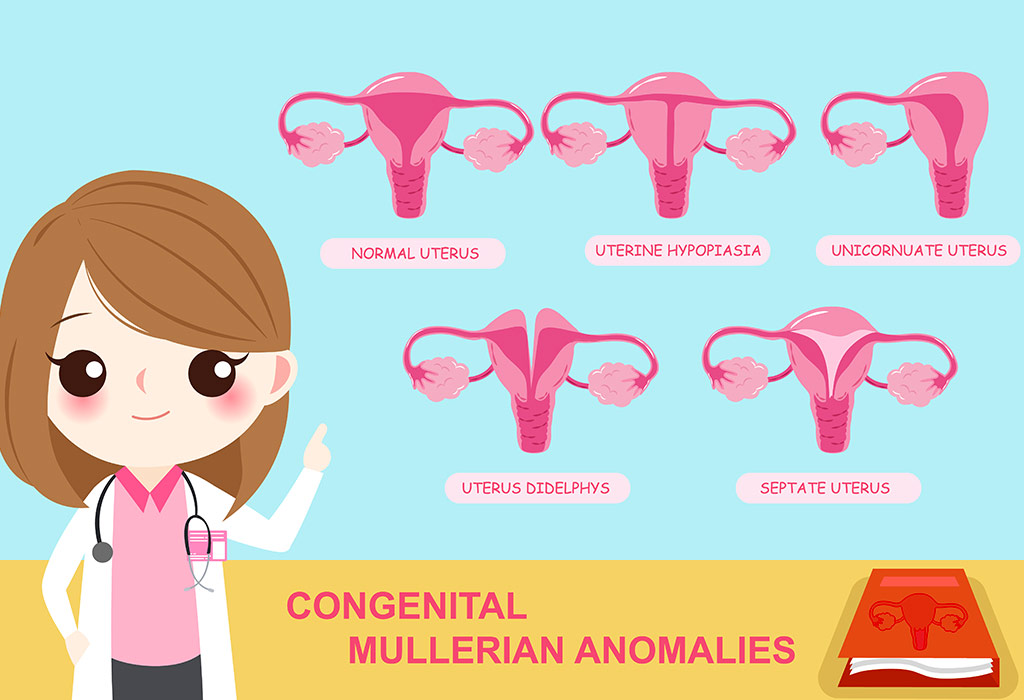
Unicornuate Uterus: Causes, Symptoms & Treatment
A unicornuate uterus is a rare congenital uterine malformation that around 0.6% of the infertile population and 0.3% of the whole population are diagnosed with. Although there have been successful pregnancies in spite of having a unicornuate uterus, there are certain risks attached to it. If you have been diagnosed with the condition and need information on it, you have reached the right place. In this article, we shall touch upon some primary points about the condition and provide insights into the causes, diagnoses and treatments to manage a unicornuate uterus to enable you to get pregnant. Read on to know more.
What Is Unicornuate Uterus?
A unicornuate uterus is a congenital uterine abnormality, in which the uterus is smaller than a typically-sized uterus and has only one functioning fallopian tube. If the other side of the uterus is present, it may be a rudimentary horn. At times, it may be connected with the rest of the uterus or may contain a functional endometrium. In certain cases, the uterus may develop a smaller offshoot called a Hemi-uterus.
What Causes Unicornuate Uterus?
A unicornuate uterus is caused due to an abnormal müllerian duct or paired duct of the embryo. It is a congenital condition, i.e. women are born with it.
Symptoms of Unicornuate Uterus
There could be no symptoms when there is a hemi-uterus with a small cavity and a functional endometrium. It is asymptomatic because the cavity forms a connection with the uterus and the vagina, and drains out the menstrual fluid. So, there is no abdominal/pelvic pain or discomfort. This occurs in 65% of women, and the condition may remain undetected until the woman faces difficulty in conceiving. In such cases, unicornuate uterus treatment becomes delayed.
But, in many cases the horn does not connect to the uterine cavity, resulting in the following symptoms:
Chronic Pelvic or Period-Related Abdominal Pain: This can occur in cases where the functioning horn is solid and isolated from the rest of the uterus. The pain occurs because of a build-up of menstrual fluid that cannot drain out through the vagina.
Hematometra: This is the accumulation or retention of blood in the uterus because of its inability to drain out fluids. The symptoms are cramping pain during periods in the lower abdomen and pelvic midline, frequent urination, and urinary retention. Women in pre-menopause may also report abnormal bleeding or no menstruation at normal cycles. The build-up of blood may also cause low blood pressure or a vasovagal issue. However, women in menopause may be asymptomatic.
A history of infertility.
Premature birth, miscarriage, and breech baby.
Diagnosis of Unicornuate Uterus
This condition remains undiagnosed until a woman suffers from pregnancy complications like a history of infertility, repeated miscarriages, or premature delivery.
There are some tests that can help detect the condition and facilitate timely treatment:
Infertility and/or Regular Pelvic Tests: Unicornuate uterus may be suspected with the help of these tests. Detection is usually rare in these tests.
Imaging: A normal ultrasound often fails to diagnose a unicornuate uterus. An MRI, which is three-dimensional sonography, has better results.
Laparoscopy: Laparoscopy can help confirm the condition by allowing a thorough check-up of the uterus.
Hysteroscopy: This is a process in which a miniature telescope is inserted via the cervix to view the uterus for diagnosis or treatment of certain conditions. This can also detect a unicornuate uterus.
Now, let’s take a look at how unicornuate uterus can affect pregnancy.
Unicornuate Uterus and Pregnancy
Reproductive complications are common in women with a unicornuate uterus. Pregnancy in unicornuate uterus women is difficult because usually only one fallopian tube functions in this situation. Even if they become pregnant unicornuate uterus involves the following risks:
Miscarriage: There maybe foetal demise and stillbirth because of the abnormal shape of the uterus and inadequate blood flow to the uterus and the placenta.
Premature Birth: Women with unicornuate uterus may go into early labour because the baby outgrows the uterine space that is smaller than usual. This happens in the first stages of pregnancy. It increases the chances of a breech birth as well, where the baby is born ‘bottom first’ instead of the normal ‘head first’. This can lead to a cesarean (C-section) instead of a normal delivery.
Ectopic Pregnancy: Women with unicornuate uterus may witness an ectopic pregnancy. This occurs when a fertilized egg implants outside the uterus, typically in the fallopian tube. Unfortunately, ectopic pregnancies must be terminated as the pregnancy cannot survive outside the uterus.
Heavy Bleeding: This could pose a life threat because of a premature rupture of membranes during the first or second trimester.
Intrauterine Growth Retardation: Here, the newborn may be smaller in weight and size.
Placenta Previa: When a woman conceives, the placenta usually attaches to the upper part of the uterus, leaving the cervix free. In placenta previa, it attaches to the lower part of the uterus, spreading to the entire or part of the cervix.
Placental Abruption: Here, the placenta gets detached from the uterus, usually after the 20th week of pregnancy. If it goes undetected, it may result in a stillborn or premature baby.
Intrauterine Foetal Demise: The condition can lead to the demise of the foetus or stillbirth.
Abdominal Pain and Collapse: Detection of the condition may be delayed in spite of the advanced technology, which could result in collapse.
With the risks mentioned above, it is clear that a unicornuate uterus affects fertility in women. Let’s find out how.
Does Unicornuate Uterus Have Any Impact on Fertility?
A unicornuate uterus causes significant gynaecological and obstetrical complications. This has a major impact on fertility. Women are rarely able to conceive when there is a rudimentary horn. Even if they manage to conceive, there are various risks involved. For example, uterine rupture during the first or the second trimester could trigger heavy bleeding and pose a threat to the mother as well as the foetus.
Here’s the rate at which the condition can affect the fertility in women:
Live birth – 29.2%
Prematurity – 44%
Ectopic pregnancy – 4%
First-trimester abortion – 24.3%
Second-trimester abortion – 9.7%
Intrauterine fetal demise – 10.5%
Although there are ways to treat fertility, treating infertility due to a unicornuate uterus may be difficult, mostly because it is a congenital condition. However, there are treatments that can allow you to manage the condition. Let’s find out how.
Unicornuate Uterus – Possible Treatments
The treatment of a unicornuate uterus is never fully effective because the uterus cannot be enlarged surgically. The surgical and non-surgical procedures given below are the most accepted methods to manage the condition:
Cervical Stitch or Cerclage: This treatment is often suggested for those cases that report a history of miscarriages, premature delivery and incompetent cervix due to a unicornuate uterus. In this procedure, the cervix is stitched and closed during pregnancy.
Laparoscopic Surgery: This treatment is conducted to remove an isolated hemi-uterus because it causes abdominal pain due to the accumulation of menstrual blood that cannot flow out.
Emergency Delivery Under Special Care: Pregnancy is possible in a non-communicating uterus, as explained earlier in the article. However, this poses greater risks to pregnant women and could involve an emergency situation because a smaller uterus may rupture by the end of the second trimester due to space constriction. Utmost care and caution should be taken to avoid risks to the mother and the baby. To avoid this, doctors recommend a laparoscopy to remove the isolated hemi-uterus.
Usually, no surgical intervention is recommended unless the endometrial tissue in the rudimentary horn causes pain or a pelvic mass. A solid non-functioning hemi-uterus also need not be removed surgically.
Finally, the type of treatment depends a lot on the variations in the unicornuate uterus.
Some of the variations are given below:
A rudimentary horn may or may not be present.
The horn may or may not connect to the rest of the uterus.
The size of the uterus can also vary.
As most conditions are not treatable, one may want to consider assisted reproduction if they have experienced one or more of the symptoms mentioned in the article above.
Unicornuate Uterus and Assisted Reproduction
One of the most accepted treatments for a unicornuate uterus is IVF with embryo transfer. eSET (elective single embryo transfer) can reduce the high risk associated with multiple pregnancies and a singleton pregnancy in women with a unicornuate uterus. If a miscarriage occurs repeatedly, surrogate motherhood can be considered.
This condition may be very complex, but the good news is that many women have given birth to full-term healthy babies. Complications can be managed and averted with careful and advanced medical care. Speak to a medical practitioner who can provide you with solutions or effective ways to manage the condition and have a baby.
References and Resources: Healthline, Verywellfamily
Also Read: List of 10 Different Types Of Pregnancy
Read more
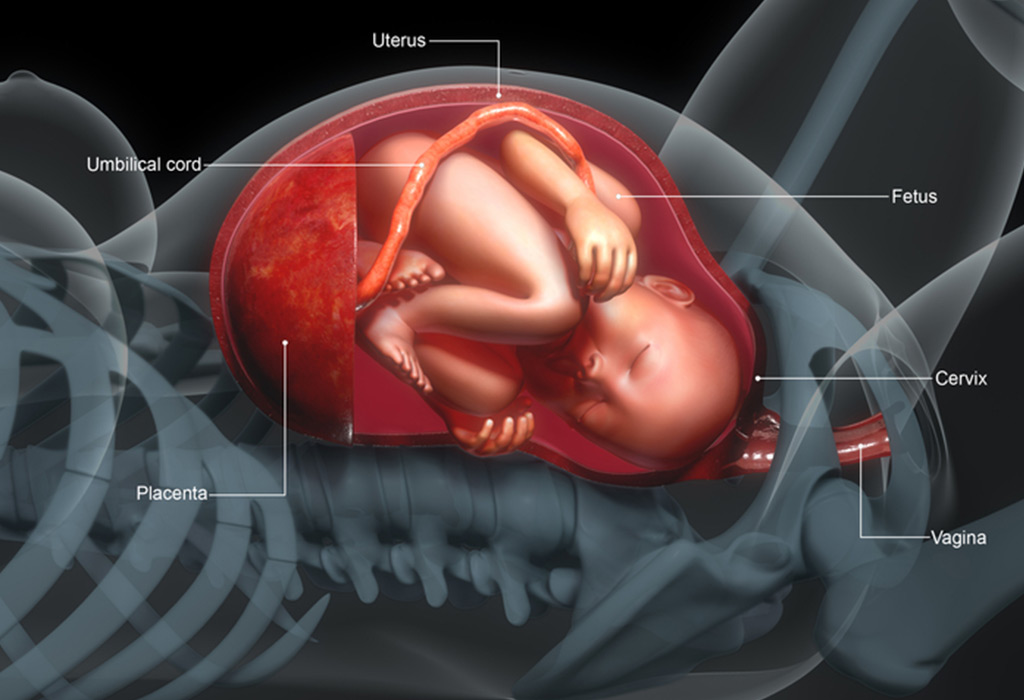
Placenta Issues in Pregnancy
The placenta is an organ that develops in a woman’s uterus when she is pregnant and provides oxygen and nutrients to the growing baby. It also filters out waste products from the baby’s blood. The placenta attaches to the wall of the uterus and umbilical cord arises from it. The placenta plays a crucial role during pregnancy. But in rare cases, the placenta may develop abnormally or be damaged, resulting in a condition called placental insufficiency. Placental insufficiency can lead to various complications and affect the health of the baby. Read about placental insufficiency in detail in this article.
What Is Placental Insufficiency?
Placental insufficiency is one of the complications that arise during pregnancy where the placenta cannot deliver enough oxygen and vital nutrients to the foetus. The other names for this condition are uteroplacental insufficiency, insufficient placenta, and placental dysfunction (1).
How Common Is Placental Insufficiency During Pregnancy?
Placental insufficiency is relatively uncommon but still affects a notable number of pregnancies. It is estimated to occur in about 3-10% of pregnancies, with higher rates in women who have conditions such as high blood pressure, diabetes, or preeclampsia (2).
What Causes Placental Insufficiency?
Some of the reasons for placental insufficiency are:
Drinking alcohol, smoking, or taking drugs during pregnancy.
Health complications such as diabetes, high blood pressure (preeclampsia), maternal blood clotting, anaemia, and if the mother is on blood thinner medications (3).
Placental abruption – a situation in which the placenta separates prematurely, before delivery (4).
In case of a placenta previa; when the placenta lies in the uterus, covering the cervix (5).
Prolonged contractions in the uterus where the blood vessels are compressed.
In case of haemorrhage due to velamentous cord insertion, where the umbilical cord is abnormally inserted into the placenta (6).
Supine hypotensive syndrome; when there is reduced blood pressure from when a mother lies down on her back, which can cause the uterus to compress the vena cava, the large veins that return blood to the heart from the head and limbs (7).
Symptoms of Placental Insufficiency
Most often there is no outward symptom of this disorder, though women who have had children before may be able to tell something is wrong as there is less movement from the child. However, in some cases, symptoms may occur and usually depend on the cause. If there is placental abruption, for example, the mother may experience pain or even notice that her uterus is extremely hard. A change in the baby’s heart rate may also be an indicator of placental insufficiency. In chronic cases, the symptoms may manifest in the infant and less in the mother. The baby may not develop properly or his heart might not work properly. Sometimes, there are no symptoms other than the sudden death of the baby (2) (3).
Diagnosis of Placental Insufficiency
Placenta insufficiency can be diagnosed during the routine ultrasound scans by monitoring the position of the placenta and by determining the small size of the uterus. Keeping a check on the alpha-fetoprotein levels and the heart rate of the baby is another way of monitoring whether or not any placental insufficiency is developing or has developed (8).
Complications Associated With Uteroplacental Vascular Insufficiency
The risks of placental insufficiency are different for both mother and child.
Risks to the Baby
The baby can suffer from deprivation of oxygen during labour. He can get hypothermia, low blood sugar and may not develop properly during pregnancy. If this disorder develops during the first trimester, it can be life-threatening to the baby. Placental insufficiency can also cause birth defects in the baby such as brain damage, lung damage, and gastrointestinal problems (2).
Risks to the Mother
A mother may have preeclampsia. If found with placental insufficiency, there is a higher chance of premature delivery, abruption in the placenta, as well as bleeding and premature contractions. There is also a higher chance she will need a C-section delivery (2). This disorder can sometimes cause post-term pregnancies, a condition where the labour gets delayed up to two weeks after the due date.
How Is Placental Insufficiency Treated?
Placental insufficiency can be treated so as to ensure as few risks as possible during the pregnancy. The approach to uteroplacental insufficiency treatment will depend on how far along a woman is into her pregnancy. For example, babies past 37 weeks in the womb are considered to be fully developed, and so the doctor may decide to induce labour or perform a C-section (2).
For cases where the condition develops and is detected early on in the pregnancy, other methods of treatment are taken into condition. Some doctors may suggest steroid shots to strengthen the baby’s lungs. Proper management of this condition will depend on further tests and the woman’s health. If the mother has diabetes or problems with blood pressure, these will need to be monitored and brought under control during pregnancy (9).
Can You Prevent Placental Insufficiency?
In order to prevent placental insufficiency in subsequent pregnancies, mothers need to ensure to get proper prenatal care. It is important for them to remain healthy and refrain from smoking, drinking and taking drugs while pregnant and even while planning for a second pregnancy.
FAQs
1. Who is at higher risk of placental insufficiency?
Women with chronic hypertension, diabetes, autoimmune disorders, obesity, or a history of pregnancy complications are at a higher risk of placental insufficiency. Smoking and advanced maternal age can also increase the likelihood of developing the condition during pregnancy (1).
2. Is it possible to have a healthy pregnancy with uterine placental insufficiency?
Yes, many women with placental insufficiency go on to have healthy pregnancies and deliveries with proper medical care and monitoring. Early diagnosis and regular prenatal checkups play an important role in improving outcomes for both the mother and the baby.
Although placental insufficiency does not endanger the life of the mother, it can affect the health of the growing baby. Special care needs to be taken and routine check-ups should not be missed as they are important to check how the baby is developing and if there’s any chance of placental insufficiency. If there is no proper management of this condition, then the baby could be born with birth problems and it could also prove to be fatal for the baby and mother. However, if detected early on and proper treatment prescribed and followed, everything should be fine!
Also Read:
Placenta Previa
Retained Placenta
Placenta in Pregnancy
Calcification of Placenta during Pregnancy
Read more

Cervical Length During Pregnancy – What Is Normal and What to Expect
Ensuring a healthy pregnancy is a top priority for expectant mothers, and understanding the various factors that may influence it is crucial. Among these factors, cervical length during pregnancy emerges as a significant consideration, as it plays a pivotal role in preventing complications during pregnancy. This article aims to provide comprehensive insights into the concept of cervical length in pregnancy and its potential impact on the gestation period. Monitoring cervical length during pregnancy is a routine part of prenatal care, and healthcare professionals often employ ultrasound examinations to assess it. According to the American College of Obstetricians and Gynecologists (ACOG), an unusually shortened cervix during pregnancy may be an indication of an increased risk of preterm labour, prompting closer monitoring and potential interventions to safeguard both the mother and the baby (1).
What Is Cervical Length?
The cervix is the lower part of the uterus that remains closed during pregnancy and gradually shortens as labour approaches. It is a three-centimetre-long canal that connects the uterus to the vagina. The cervical length is simply the length of the canal, including the cervical opening. In pregnant women, the cervix needs to be of a specific length for the growth of a healthy baby. Measuring cervical length helps doctors assess the risk of preterm birth, especially in women with a history of preterm delivery or other risk factors.
Why Is the Cervical Length Important?
The cervix is almost inflexible until you get pregnant. Once you conceive, it becomes softer and shorter, loses muscle tone, and dilates. This is known as cervical effacement, which allows the foetus to fit inside the cervical canal while being born. However, if your cervix is too short, you may be at risk of preterm labour and all the complications that come along with premature birth (2).
Normal Cervical Length During Pregnancy Chart
A normal cervical length in pregnancy varies slightly throughout the terms, but it generally remains above 25 mm (2.5 cm) until later gestation. The following chart provides approximate normal ranges commonly used in obstetric practice.
Week
Normal Cervical Length
12 – 14 weeks
35–50 mm
16–18 weeks
35–50 mm
20–24 weeks
35–50 mm
24–28 weeks
30–45 mm
28–32 weeks
30–40 mm
32–36 weeks
25–40 mm
36–40 weeks
Gradually shortens in preparation for labour
Are Having a Short Cervix and Cervical Insufficiency Related?
No, a short cervix and cervical insufficiency are totally different things. A short cervix merely refers to the length of the cervical canal. Cervical insufficiency, on the other hand, refers to the premature cervical effacement and dilation that happen when the muscles are unable to remain firm and strong. Cervical insufficiency can lead to a short cervix.
How Do I Know If My Cervix Is Short?
A short cervix often causes no symptoms and is usually detected during an ultrasound examination. However, some women may experience:
Increased pelvic pressure
Persistent lower backache
Mild abdominal cramping
Changes in vaginal discharge
Light spotting or bleeding
Sensation that the baby is pushing downward
Can Cervical Length Change During Pregnancy?
Yes. Cervical length naturally changes throughout pregnancy. In most women, the cervix remains long and closed until the third trimester. However, some women may experience earlier shortening, which can increase the risk of preterm birth. Regular ultrasound monitoring may be recommended for those at higher risk.
Factors That Influence the Cervical Length
Several factors influence cervical length. Some of them are:
1. Anatomy
Some women have a short cervix. That’s how their bodies are built.
2. Distended Uterus
In multiple pregnancies, the uterus stretches beyond its capacity. It pushes downwards on the cervix, reducing its length.
3. Cervical Insufficiency
Cervical insufficiency or an incompetent cervix is a condition caused by the lack of cervical muscle strength, which can influence cervical length (3).
How Is the Length of the Cervix Measured?
An ultrasound transvaginal scan can help to measure the cervical length. Ideally, the cervical length in the 24th week should be about 3.5 – 5 cm, whereas at 28 weeks, it should be 3.5 – 4 cm. At 32 weeks, the cervical length for normal delivery in cm is 3 – 3.5 cm. A cervix shorter than 2.5 cm increases the likelihood of preterm birth.
Is Measuring the Cervical Length a Must?
Doctors usually recommend the cervical length scan to women carrying twins or multiples or having a medical history of premature birth and miscarriage.
What Are the Risks Associated With Short Cervical Length?
Preterm birth stands out as a significant risk associated with a short cervical length (4). Beyond potential health challenges for the infant, premature delivery elevates the likelihood of complications such as preeclampsia, haemorrhage, and infection for the mother.
It’s essential to recognise that a shortened cervix can also be a contributing factor to cervical incompetence, characterised by premature cervical opening typically occurring in the second trimester. Cervical incompetence poses a severe threat, potentially leading to miscarriage or preterm birth if not addressed promptly.
Moreover, a reduced cervical length heightens the probability of requiring a cesarean delivery. This is attributed to the potential inadequate dilation of the shortened cervix during labour, creating difficulties for the baby to traverse the birth canal. In certain instances, opting for a cesarean delivery becomes imperative to ensure the safety and well-being of both the mother and the newborn.
Tips to Deal With a Short Cervix
Here are a few things you should do if your cervix is too short:
1. Bed Rest
Bed rest shouldn’t be compromised. Lying down prevents the uterus and the foetus from bearing down on the cervix, thus preventing it from early effacement.
2. Cerclage
This is a minor surgery in which your doctor will stitch your cervix shut with a couple of sutures. The procedure is quick and performed under anaesthesia. It is done for women who have had prior preterm deliveries. Some doctors believe that cerclages can lead to infection and inflammation, but these can be controlled with the right medications.
3. Hormonal Treatment
In this treatment, your doctor will place the pregnancy hormone, progesterone, in the cervical area. Progesterone can help in the relaxation of the uterus.
4. Pessary
A silicone device known as a pessary can be used to support the cervix, keeping it closed shut.
Cervical Length in Twin or Multiple Pregnancies
Women expecting twins or triplets are more likely to experience early labour (spontaneous preterm birth) compared to those who are pregnant with a single baby (5). While normal cervical length varies, the average mid-pregnancy CL for twin gestations is typically between 35 mm and 40 mm (6). To give you a context on when the cervical length remains in the normal range and when it exceeds (7):
Low Risk: Generally ≥ 30 mm.
Borderline/Close Monitoring: Below 30 mm to 35 mm.
High Risk (Short Cervix): ≤ 25 mm before 24 weeks of gestation is often used as an indicator to signal a high risk for spontaneous preterm delivery.
When to Consult a Doctor?
It is important to talk to your doctor about any worries regarding cervical length, particularly if you notice:
Any indications or symptoms of a shortened cervix.
A background of preterm labour or cervical insufficiency.
Risk factors for cervical shortening include multiple pregnancies or past cervical procedures.
FAQs
1. Can Cervical Length Be Improved During Pregnancy?
Cervical length in pregnancy is generally a biological characteristic and cannot be significantly altered. However, certain interventions, like progesterone supplementation or cervical cerclage, may be recommended by healthcare professionals in specific cases to address potential issues.
2. How Often Should Cervical Length Be Monitored During Pregnancy?
The frequency of cervical size during pregnancy monitoring varies based on individual risk factors and healthcare provider recommendations. Generally, healthcare professionals assess cervical length during routine ultrasound examinations, with more frequent monitoring for those at higher risk of complications.
3. What Can I Do to Maintain a Healthy Cervical Length During Pregnancy?
While cervical length is largely influenced by biological factors, maintaining overall health is essential. Following a balanced diet, staying hydrated, avoiding tobacco and illicit substances, and attending regular prenatal check-ups contribute to overall well-being. However, specific interventions for cervical length are typically determined and guided by healthcare providers based on individual circumstances.
It’s important to understand the changes your body goes through during pregnancy. Cervical length plays a major role during pregnancy, so it is essential to stay informed about it. Do not worry if you have a normal cervical length in pregnancy. If you have any concerns about the cervical length during pregnancy, consult your gynaecologist.
Also Read:
Cervical Position in Pregnancy
Can you get Pregnant with Small Uterus
Cervix Dilation during Labour and Birth
Read more
Sort By
 Latest
 Most Popular
 Trending
Please select atleast one Filter