


16 Weeks Pregnant Ultrasound : What to Expect?
Your 16 week ultrasound checks fetal growth, heartbeat and organ development. Know what the scan involves and how to prepare.




In this Article
- Why Should Pregnant Women Have an Ultrasound at 16 Weeks?
- What Preparation Is Needed for a 16-week Scan?
- How Long Does a Sixteen-Week Ultrasound Take to Complete?
- How Is Sixteen Week Scan Done?
- What Can Be Seen in the Scan?
- Is It Possible to Determine the Gender of the Baby at a 16 Week Ultrasound?
- What If Any Abnormalities Are Seen in the Ultrasound?
- FAQs
View More
Hey, it’s your sweet 16th month of pregnancy! You are beginning your fourth month and entering the second trimester. There’s a lot happening inside your body, and your doctors mean well for you if they suspect anything serious. About 1 in 100 babies is born with a heart defect. Don’t worry; early interventions, like a 16-week ultrasound, can spot issues early. While 16-week ultrasounds aren’t routine, the possibility of a heart defect isn’t just one reason your doctor would recommend you have the 16-week scan. If you are scheduled for one, you will likely have it transabdominally to help the doctor assess the baby’s development, including growth of the torso, limbs, head, and facial features. In this article, we discuss everything crucial you need to expect from your ultrasound at 16 weeks. Read on!
Why Should Pregnant Women Have an Ultrasound at 16 Weeks?
Things start to get a bit exciting once you hit the 16-week mark, and an ultrasound is important to understand your baby’s progress. Prenatal care is an integral part of every mother’s journey, and here are a few reasons as to why an ultrasound is necessary at this stage.
- It can help you understand your baby’s growth, and the sonographer can help measure certain parts of your baby’s body to assess any concerns.
- Ultrasound will become an integral part of your journey into motherhood. It helps determine the size, structure and any pathological lesions on the foetus (1).
- It can present a visual representation of your baby and help prepare for routine and emergency prenatal care.
- It can help understand if your baby is healthy and safe and if there are any abnormalities.
- Genetic issues can also be determined, such as Down Syndrome or any other chromosomal defects, or have any other neural tube defect.
- The 16-week foetus ultrasound can determine the heartbeat and health of the heart and other organs. It can help determine if there is any heart defect as well.
What Preparation Is Needed for a 16-week Scan?
Your baby at 16 weeks’ ultrasound is the largest it has been in your pregnancy to date, and a few things need to be done before you go for a 16-week scan. Your baby, which measures around four and a half inches during the 16th week, will grow double in size very quickly, month by month, from here onwards. There will definitely be a weight gain during this time, but morning sickness will completely disappear.
Your body will start producing milk, which will lead to breast augmentation. In addition, the belly will be larger, and the skin colour will change to some extent as well.
Before a scan, it is important to carry documents about your and your partner’s medical history to your gynaecologist and ask questions regarding any symptoms or issues you may be experiencing. You should also get a full physical examination, including thyroid, breasts, lungs and heart. You should get a lab test done to check signs of gestational diabetes, preeclampsia or any other urinary tract infections through urine tests.
Familiarising oneself with terms which are associated with pregnancy and medical procedures, such as ultrasound, will also help in understanding your baby better.
How Long Does a Sixteen-Week Ultrasound Take to Complete?
An ultrasound can last anywhere between 30 and 60 minutes. Before an ultrasound, a patient is asked to drink lots of water or fluids and stay away from solid food. You cannot access the toilet when you wait for the scan, but you can go relieve yourself once the procedure is complete (2).
Before you begin the test, make sure to prepare yourself mentally to wait and to understand that they take time, to image the sonography. Imaging reports also take a day to be produced, but both parents will be shown the image of their baby on the screen.
How Is Sixteen Week Scan Done?
A 16-week foetus ultrasound is done by using a device that emits high-frequency sound waves into the body. The waves hit and bounce off the various organs of the body as they travel. A probe picks up these echoes created by the bouncing sound waves and starts converting them into a moving image. These images can be accessed through a graphics display monitor. These tests are usually conducted in clinics, diagnostic centres or the radiology section of a hospital. A specially trained expert will be the only one who will be able to conduct a scan.

What Can Be Seen in the Scan?
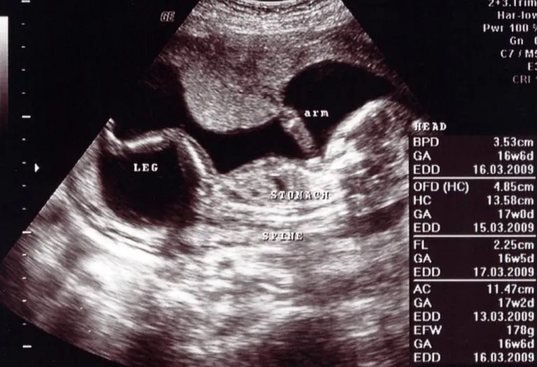
When you get an ultrasound done at 16 weeks, your belly will look like a small cantaloupe. On the inside, however, your baby is around 4-5 inches in size and will continue to grow. Hair and fingernails are starting to develop, although they will be more noticeable once you progress in your pregnancy. You will be able to make out the head, the eyes, ears and torso of your baby. All the muscles and bones are in place, and the central nervous system is starting to exert control, so you may even see your baby move during an ultrasound.
Your baby is beginning to make expressions, but any smiling or frowning will happen randomly, as they lack muscle control at this stage. The nervous system is still maturing, allowing your baby to start moving their arms and legs. Your baby’s hands can clench into fists, and they might start jabbing around inside you as well (3).
Is It Possible to Determine the Gender of the Baby at a 16 Week Ultrasound?
Yes, you can ascertain your baby’s gender during the 16-week gender scan. The determination should be reliable since the baby’s external anatomy is fully developed by then.
However, it’s important to note that accuracy may be compromised by the baby’s positioning, potentially hindering the physician or ultrasound technician from obtaining a clear view of the baby’s anatomy for gender confirmation.
Disclaimer: Under the Pre-conception and Pre-natal Diagnostic Techniques (Prohibition of Sex Selection) Act, 1994, (PC & PNDT Act), determining or disclosing the sex of a fetus before birth is illegal in India, except for specific medical indications permitted by judicial law. Therefore, all healthcare providers, including doctors, clinics, and hospitals, in India cannot reveal a baby’s gender during a 16-week fetus ultrasound or at any stage of pregnancy (4).
What If Any Abnormalities Are Seen in the Ultrasound?
When you go for an ultrasound, the sonographer will also conduct a variety of other tests to determine any abnormalities in your baby’s growth such as measuring the length of the baby’s nasal bone to determine any abnormalities in growth is one. There are several blood tests which are offered to determine any risks or abnormalities that can arise at this stage of the pregnancy.
The Triple screen is a prenatal blood test that will help assess any issues with the baby. The test primarily helps test levels of alpha-fetoprotein (AFP), a protein produced by the foetus’s liver and two hormones: human chorionic gonadotropin (hCG) and estriol, to determine any birth defects such as Down Syndrome, Spina Bifida (5).
It is important to discuss any concerns or anything that comes up during an ultrasound with your doctor in order to determine the next course of action for you and your baby. It is also important to take any medication or tests that are required to keep you and your baby healthy.
FAQs
1. Is a 16-week ultrasound safe for my baby?
Yes. Diagnostic ultrasounds use sound waves rather than radiation and are considered safe when performed by trained healthcare professionals for medical purposes. Thus, an ultrasound in a 16-week pregnancy is safe.
2. Can I expect twins at a 16-week ultrasound?
Yes, a normal 16-week ultrasound can show babies in detail if you are carrying twins. You can later have a 16-week twin ultrasound to confirm.
3. Which test can be done at 16 weeks?
Between 16 and 18 weeks of pregnancy, your doctor may provide you with a second-trimester screening test (16 week 3d ultrasound or normal ultrasound), known as the multiple marker test or triple screen.
4. Can a 16-week ultrasound detect birth defects?
Some structural abnormalities may be visible at 16 weeks, but the detailed anatomy scan performed around 18–22 weeks is usually more effective for identifying congenital conditions.
At 16 weeks every mother will have the pregnancy glow and move into the second trimester which is an enjoyable phase. As your baby continues to grow, more movement from the foetus will show, the belly will grow bigger, and the body prepares for the next phase of motherhood.
Previous Week: 15 Weeks Pregnant Ultrasound
Next Week: 17 Weeks Pregnant Ultrasound
Was This Article Helpful?
Parenting is a huge responsibility, for you as a caregiver, but also for us as a parenting content platform. We understand that and take our responsibility of creating credible content seriously. FirstCry Parenting articles are written and published only after extensive research using factually sound references to deliver quality content that is accurate, validated by experts, and completely reliable. To understand how we go about creating content that is credible, read our editorial policy here.
- Author


Related Articles
-

Excessive Hair Growth During Pregnancy - Is It Normal?
-

Gallstones in Pregnancy - Symptoms, Causes, Treatment & Prevention
-

What To Do & Not To Do in Your Third Trimester of Pregnancy?
-

Acrylic Nails During Pregnancy – Is It Safe?
-

Baby Positions in the Womb and How to Tell them
-

10 Healthy Ways to Keep Karva Chauth Fast When Pregnant