8 Weeks Ultrasound – What to Expect
Learn what happens at an 8 weeks ultrasound, from baby’s growth to heartbeat and early pregnancy checks

- Why Should You Have an Ultrasound Scan at 8 Weeks?
- How to Prepare for Your Eighth-Week Scan
- How Long Does the 8 Week Sonography Take?
- Ultrasound Picture at 8 Weeks Pregnant
- What Can Your Doctor Determine?
- What Can You Expect to See at the Eighth Week Ultrasound?
- Is It Possible to Miss Twins on 8-Week Ultrasound?
- If There’s No Heartbeat at 8 Weeks, Can It Be a Miscarriage?
- Is It Possible to Identify the Gender of the Baby at 8th-Week Ultrasound?
- What If Any Other Abnormalities Are Detected During 8 Week Ultrasound?
- FAQs
In your pregnancy journey, you embark on a miraculous journey where nature weaves its delicate tapestry, nurturing a tiny life. An ultrasound scan gives you a glimpse of your growing baby. The thought of your first ultrasound can make you both anxious and excited. Timely ultrasounds also help the gynaecologists gauge the health of the baby inside the womb and confirm a healthy pregnancy. If you are nearing the 8th week of your pregnancy, you must be anxious about the upcoming 8 weeks ultrasound in pregnancy. Well, don’t be. In this article, we will share everything you need to know about an ultrasound scan at 8 weeks. Let’s begin with why you need the 8 week sonogram.
Why Should You Have an Ultrasound Scan at 8 Weeks?
An ultrasound at eight weeks isn’t a must. However, your gynaecologist may suggest a scan to check your foetus’ health. This ultrasound scan may be performed for the following reasons (1):
- To get an idea about the gestational age of your baby.
- To determine the cause of bleeding, if any.
- To check for multiple pregnancies.
- To check whether your baby has a heartbeat.
- To ascertain the size of the embryo.
- To check the health of the fallopian tubes and the ovaries.
- To check for any complications such as an ectopic pregnancy.
How to Prepare for Your Eighth-Week Scan
An abdominal scan or a transvaginal scan may be performed during the 8th week. Both scans have slightly different requirements, though.
An abdominal scan requires you to have a full bladder. As you are in the initial few weeks of pregnancy, your baby will be very small. So, a full bladder is required to push the uterus up. This helps in obtaining a better image of your baby. Wear loose and comfortable clothing, as you may have to expose your tummy for the scan. In this type of scan, a conductive gel is spread over the belly, and a handheld scanner is run over it. The gel enables ultrasound waves to pass to the uterus. The waves then bounce back, giving a glimpse of the foetus.
On the other hand, for a transvaginal ultrasound scan, the bladder should be empty. A full bladder may distort the visuals. In this type of scan, a probe is inserted inside the vagina. It is pressed against the cervix to obtain clear pictures. At times, you might feel slight pressure (2).

How Long Does the 8 Week Sonography Take?
Generally, the ultrasound scan takes around 20 to 30 minutes. However, it might take longer if the foetus is oddly positioned. Sometimes, dense tissues may also make it difficult and time-consuming to obtain clear images of the baby.
Ultrasound Picture at 8 Weeks Pregnant
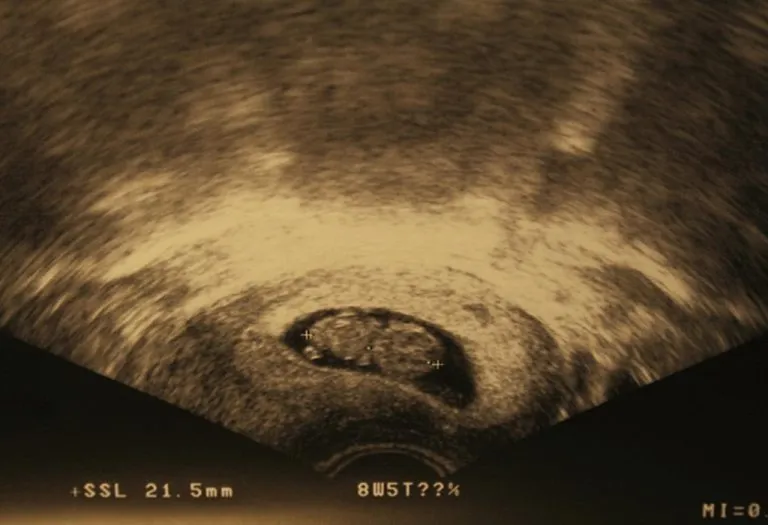
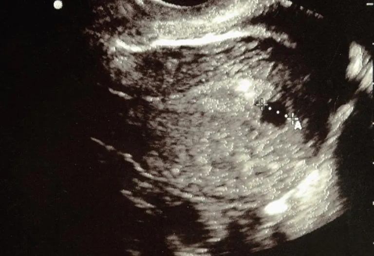
What do 8-week ultrasounds look like? Your ultrasound won’t produce a sharp, colour picture. Instead, you’ll get a blurry black-and-white image. Despite that, it’s such a special glimpse of your baby that you’ll likely display it proudly. Here is an example of what an 8-week sonogram picture often looks like.
What Can Your Doctor Determine?
If you’re wondering what you can expect to see in an ultrasound scan at 8 weeks, we have the answer! Here’s what your doctor can determine with this scan (3):
- Size of the foetus.
- The presence of the baby’s heartbeat and the rate at which her heart is beating.
- The presence of multiple babies can be determined by the presence of multiple heartbeats and multiple yolk sacs.
- Vague images of the growth of tiny hands, legs, and the formation of eyes, nostrils, internal organs, and the mouth.
What Can You Expect to See at the Eighth Week Ultrasound?
This is what the embryo looks like at 8 weeks (4):
- The eyes would have migrated from the sides to the front
- The embryo would measure 2.3 cm from crown to rump
- The outer ears would be starting to form
- Your baby can move his elbows
- The fingers look like tiny buds
- The digestive system would have started developing
- The baby’s head still bends over his stomach
Is It Possible to Miss Twins on 8-Week Ultrasound?
Yes, it is possible to miss the presence of twins during an 8-week ultrasound. Although ultrasound technology is generally reliable, certain circumstances may lead to the oversight of twin pregnancy at this early stage. Factors such as the position of the embryos, the skill of the sonographer, and the clarity of the ultrasound image can influence the diagnosis’s accuracy.
About the 8-week ultrasound twins, the embryos are quite small, and their distinct features may not yet be fully visible. Additionally, if the embryos are positioned close together or overlap, it can be challenging to distinguish them as separate entities. In some cases, the sonographer may inadvertently focus on one embryo and overlook the presence of another. It is important to note that the likelihood of detecting twins increases as the pregnancy progresses and subsequent ultrasounds are conducted. As the embryos grow and develop, their individual structures become more pronounced, making it easier to identify multiple pregnancies.
If There’s No Heartbeat at 8 Weeks, Can It Be a Miscarriage?
In some of the cases, you may not be able to see or hear the heartbeat at 8 weeks. However, that doesn’t confirm a miscarriage. Your gynaecologist may suggest a follow-up scan in the forthcoming week to rule out a miscarriage in an 8-week ultrasound.
A miscarriage has other symptoms, too, so the absence of a heartbeat shouldn’t make you jump to conclusions.
Is It Possible to Identify the Gender of the Baby at 8th-Week Ultrasound?
At the 8th week of pregnancy, it is generally not possible to identify the gender of the baby through ultrasound. During this early stage, the sexual organs of the foetus are still in the early stages of development and may not be distinguishable on the ultrasound image.
The formation of the external genitalia typically occurs around 11 and 14 weeks of pregnancy. Therefore, a more accurate determination of the baby’s gender can usually be made during the anatomy scan, typically conducted around the 18th to 20th week of pregnancy. At this stage, the genitals are more developed and identifiable, allowing the sonographer to determine the gender of the baby with greater accuracy potentially (5).
Even during later ultrasounds, determining the baby’s gender is not always 100% guaranteed. Factors such as the baby’s position, amniotic fluid levels, and the sonographer’s skill can influence the assessment’s accuracy. For a definitive confirmation of the baby’s gender, additional diagnostic methods, such as genetic testing or amniocentesis, may be recommended by a healthcare professional.
What If Any Other Abnormalities Are Detected During 8 Week Ultrasound?
During an 8-week ultrasound, various abnormalities or concerns may be detected, although it is important to note that the ultrasound at this stage is primarily performed to assess the viability and early development of the pregnancy.
Here are a few examples of abnormalities that may potentially be identified:
- Miscarriage or ectopic pregnancy
- Gestational age discrepancy
- Multiple pregnancies
- Abnormalities in the gestational sac or yolk sac
- Fetal heartbeat concerns
FAQs
1. Is it possible to have an 8-week ultrasound in 3D?
It is less common to have a 3D ultrasound at 8 weeks. Typically, 3D ultrasounds are performed later in pregnancy when the baby’s features are more developed and recognisable. At 8 weeks, the embryo is still in the early stages of development, and the image produced by a traditional 2D ultrasound is usually sufficient for assessing the pregnancy.
2. Can I take my partner to the 8-week scan?
Usually, having your partner accompany you to the eight week ultrasound is generally allowed and encouraged. The early ultrasounds are important moments for both parents to witness the initial stages of pregnancy and share the joy and anticipation of seeing the baby’s development. However, checking with your healthcare provider or the facility where the ultrasound will be conducted is recommended to confirm their specific policies regarding accompanying guests.
3. How does the size and image of the foetus change from 8 to 9 weeks?
From 8 to 9 weeks, the fetus undergoes rapid growth and development. During this time, the size of the fetus increases as it gains weight and its features become more defined. By 9 weeks, the embryo has developed into a fetus, and its body proportions resemble a human’s. Additionally, the ultrasound image at 9 weeks may provide clearer details of the fetus, including the head, limbs, and internal organs, compared to the earlier 8-week gestation ultrasound. However, it’s important to note that specific changes and variations can differ among pregnancies, and individual factors may influence the appearance and growth rate of the fetus.
4. The ultrasound picture shows a large, empty black circle near the baby. Should I be worried?
Not at all! That large black circle is almost certainly the gestational sac, which is the fluid-filled sac that surrounds and protects your developing baby. It’s supposed to be there and is a very important and reassuring structure. In fact, one of the first things the sonographer checks is that this sac is present and in the right location.
If any abnormalities or concerns are detected during an 8-week embryo ultrasound, further evaluations and discussions with a doctor are typically recommended. Based on the specific findings and circumstances, they will provide appropriate guidance, additional testing, and necessary support.
An ultrasound scan is more like a camera taking a picture of your growing baby. However, it’s good to remember that every baby is different, and so the ultrasound scan reports vary from baby to baby. When in doubt, do not fret, but consult your doctor.
Previous Week: 7 Weeks Pregnant Ultrasound
Next Week: 9 Weeks Pregnant Ultrasound
Was This Article Helpful?
Parenting is a huge responsibility, for you as a caregiver, but also for us as a parenting content platform. We understand that and take our responsibility of creating credible content seriously. FirstCry Parenting articles are written and published only after extensive research using factually sound references to deliver quality content that is accurate, validated by experts, and completely reliable. To understand how we go about creating content that is credible, read our editorial policy here.


